Copyright
©The Author(s) 2019.
World J Clin Cases. Jan 26, 2019; 7(2): 191-202
Published online Jan 26, 2019. doi: 10.12998/wjcc.v7.i2.191
Published online Jan 26, 2019. doi: 10.12998/wjcc.v7.i2.191
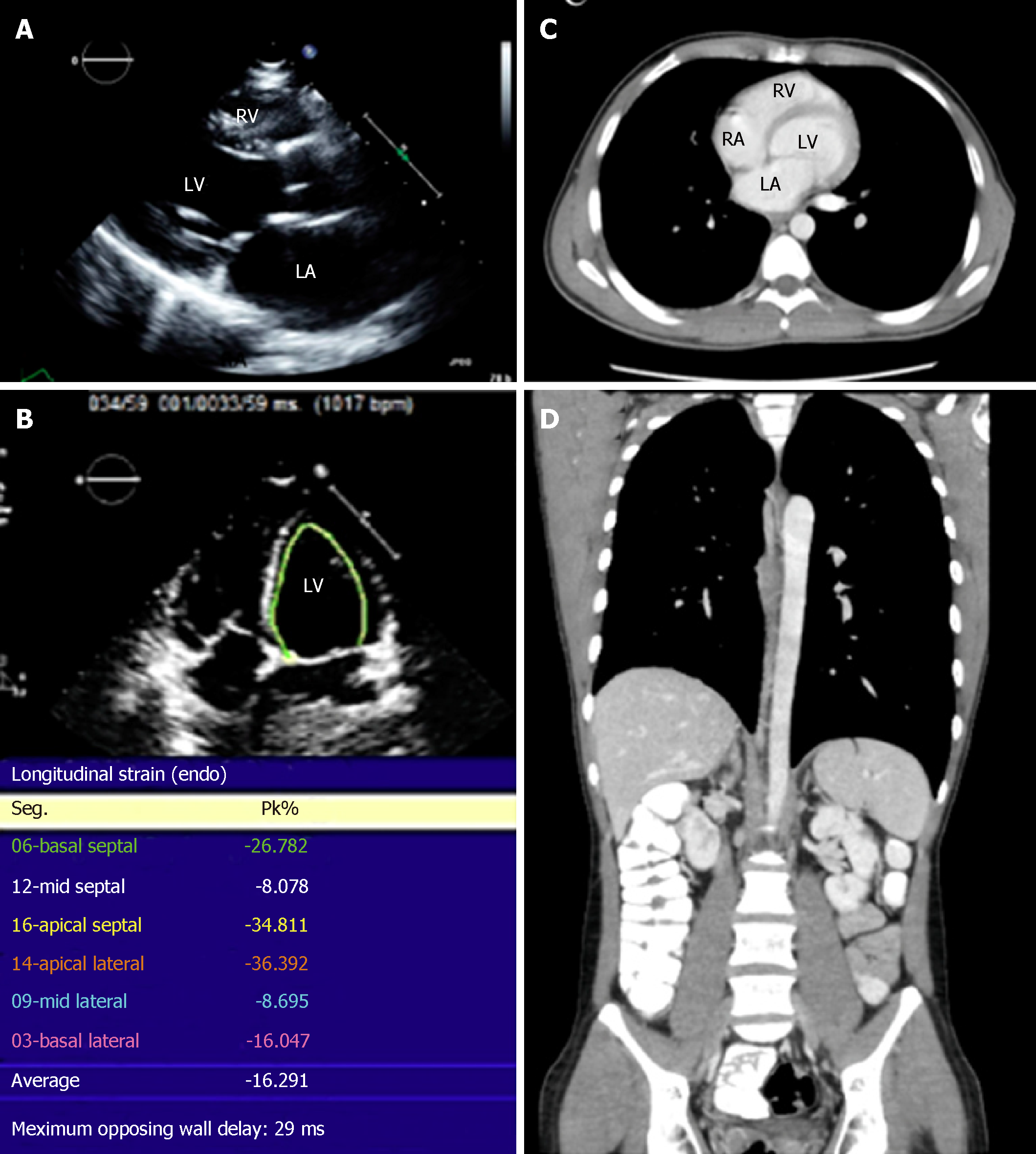
Figure 3 Recent imaging of the patient showing resolution of the lymphoma.
A: Normal parasternal long axis echocardiographic view; B: Strain imaging in a 4-chamber view showing improvement in lateral wall strain; C and D: Normal computed tomography images of the patient; C: Axial image of the chest; D: Coronal thoracoabdominal image. RV: Right ventricle; LV: Left ventricle; RA: Right atrium; LA: Left atrium.
- Citation: Al-Mehisen R, Al-Mohaissen M, Yousef H. Cardiac involvement in disseminated diffuse large B-cell lymphoma, successful management with chemotherapy dose reduction guided by cardiac imaging: A case report and review of literature. World J Clin Cases 2019; 7(2): 191-202
- URL: https://www.wjgnet.com/2307-8960/full/v7/i2/191.htm
- DOI: https://dx.doi.org/10.12998/wjcc.v7.i2.191


