Copyright
©The Author(s) 2019.
World J Clin Cases. Oct 6, 2019; 7(19): 3145-3152
Published online Oct 6, 2019. doi: 10.12998/wjcc.v7.i19.3145
Published online Oct 6, 2019. doi: 10.12998/wjcc.v7.i19.3145
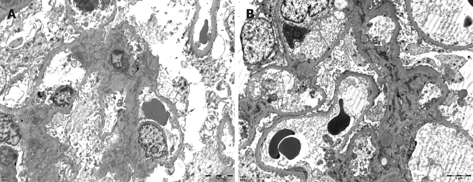
Figure 3 Renal pathology of nephrotic syndrome by electron microscopy.
After toluidine blue staining, two glomeruli were observed. One of them was observed by ultrathin section of glomerulus under an electron microscope. Erythrocytes aggregated in glomerular capillary lumen, capillary endothelial cells did not proliferate, and capillary loops were open. No obvious thickening of the wall of renal capsule, vacuolar degeneration of the wall cells, or proliferation of the wall cells was observed. Basement membrane showed no obvious thickening, with a thickness of about 300-450 nm. Visceral epithelial cells showed swelling, vacuolar degeneration, and diffuse fusion of foot processes. In the mesenchymal area, mesenchymal cells and matrix proliferated slightly, no exact electron dense deposits were found, and no electron dense deposits were found under the epithelium and endothelium. Vacuolar degeneration of renal tubular epithelial cells was noted, without special pathological changes in renal interstitium. Erythrocyte aggregation was seen in the lumen of individual renal interstitial capillaries. A diagnosis of focal segmental glomerulosclerosis was made (bar = 0.5 μm).
- Citation: Wu J, Li P, Chen Y, Yang XH, Lei MY, Zhao L. Hypereosinophilia, mastectomy, and nephrotic syndrome in a male patient: A case report. World J Clin Cases 2019; 7(19): 3145-3152
- URL: https://www.wjgnet.com/2307-8960/full/v7/i19/3145.htm
- DOI: https://dx.doi.org/10.12998/wjcc.v7.i19.3145


