Copyright
©The Author(s) 2019.
World J Clin Cases. Oct 6, 2019; 7(19): 3145-3152
Published online Oct 6, 2019. doi: 10.12998/wjcc.v7.i19.3145
Published online Oct 6, 2019. doi: 10.12998/wjcc.v7.i19.3145
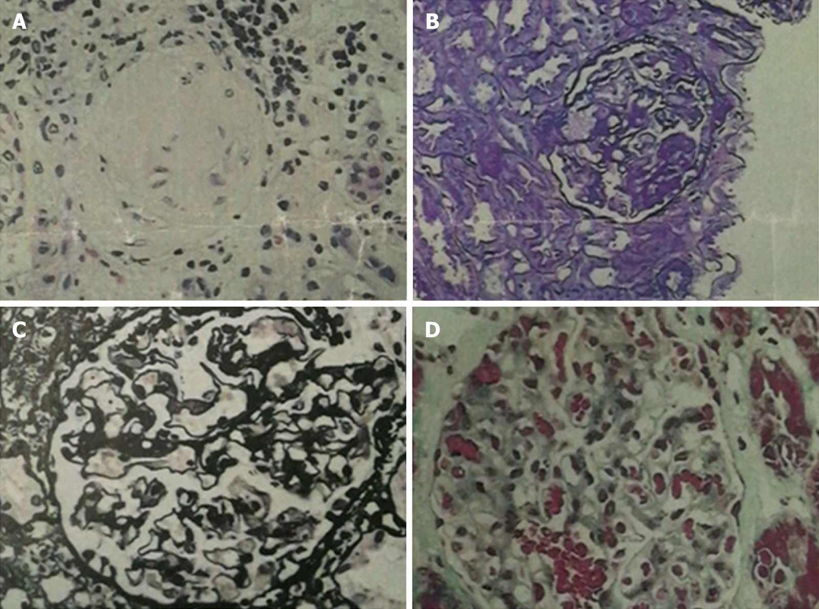
Figure 2 Renal pathology of nephrotic syndrome by analysis of morphological pattern using different staining methods.
The specimens were sliced at three layers, mainly in the renal cortex. The number of glomeruli at three layers was 23, 28, and 25, respectively. Glomerular sclerosis in four glomeruli, and glomerular segmental sclerosis and podocyte proliferation in one were found in 28 glomeruli of the second layer. The glomerular mesangial cells and matrix proliferated slightly, the capillary loops opened, and the basement membrane did not thicken. There was no obvious deposition of fumoglobin in the mesangium, subepithelium, or subendothelium, and no mesangial insertion or double track formation. No proliferation of parietal cells and no crescent formation were noted. The epithelial cells of renal tubules showed vacuolar degeneration, focal atrophy (about 5%), infiltration of small focal inflammatory cells in the renal interstitium, thickening of small arterial wall, and narrowing of lumen. Under fluorescence, IgM (+), IgA, IgG, C3, and C1q (-), and C3 (+) of arterioles were observed. The diagnosis was focal segmental glomerulosclerosis, non-specific type (FSGS, NOS). A: Hematoxylin-eosin stained (×400) histological image showing a sclerotic glomerulus; B: Periodic acid-Schiff stained (×400) histological image showing segmental glomerulosclerosis; C: Periodic acid-Schiff methenamine stained (×400) histological image showing open capillary loops and no significant thickening of basement membrane; D: Masson stained (×400) histological image showing no significant deposition of hemophilic protein.
- Citation: Wu J, Li P, Chen Y, Yang XH, Lei MY, Zhao L. Hypereosinophilia, mastectomy, and nephrotic syndrome in a male patient: A case report. World J Clin Cases 2019; 7(19): 3145-3152
- URL: https://www.wjgnet.com/2307-8960/full/v7/i19/3145.htm
- DOI: https://dx.doi.org/10.12998/wjcc.v7.i19.3145


