Copyright
©The Author(s) 2019.
World J Clin Cases. Oct 6, 2019; 7(19): 3145-3152
Published online Oct 6, 2019. doi: 10.12998/wjcc.v7.i19.3145
Published online Oct 6, 2019. doi: 10.12998/wjcc.v7.i19.3145
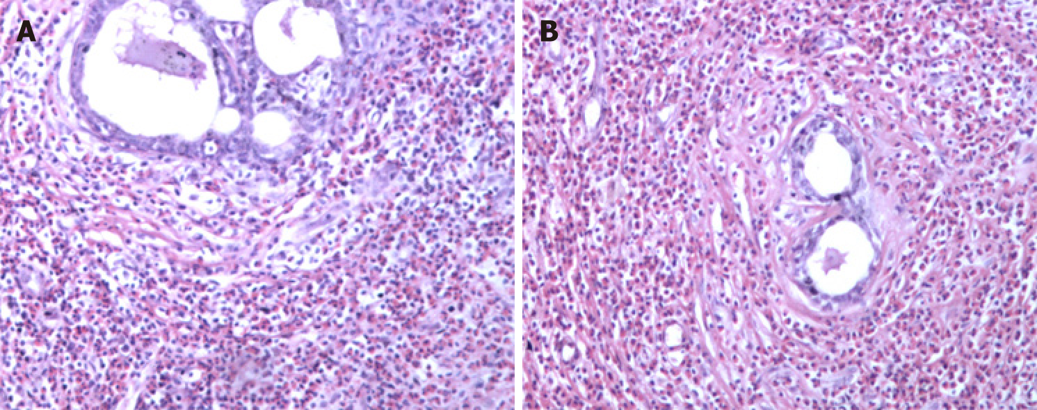
Figure 1 Breast pathology showing breast involvement by analysis of cell morphology (hematoxylin-eosin staining, ×100).
A and B: Intra-tissue ductal dilatation and interstitial eosinophil infiltration are shown.
- Citation: Wu J, Li P, Chen Y, Yang XH, Lei MY, Zhao L. Hypereosinophilia, mastectomy, and nephrotic syndrome in a male patient: A case report. World J Clin Cases 2019; 7(19): 3145-3152
- URL: https://www.wjgnet.com/2307-8960/full/v7/i19/3145.htm
- DOI: https://dx.doi.org/10.12998/wjcc.v7.i19.3145


