Copyright
©The Author(s) 2019.
World J Clin Cases. Oct 6, 2019; 7(19): 3138-3144
Published online Oct 6, 2019. doi: 10.12998/wjcc.v7.i19.3138
Published online Oct 6, 2019. doi: 10.12998/wjcc.v7.i19.3138
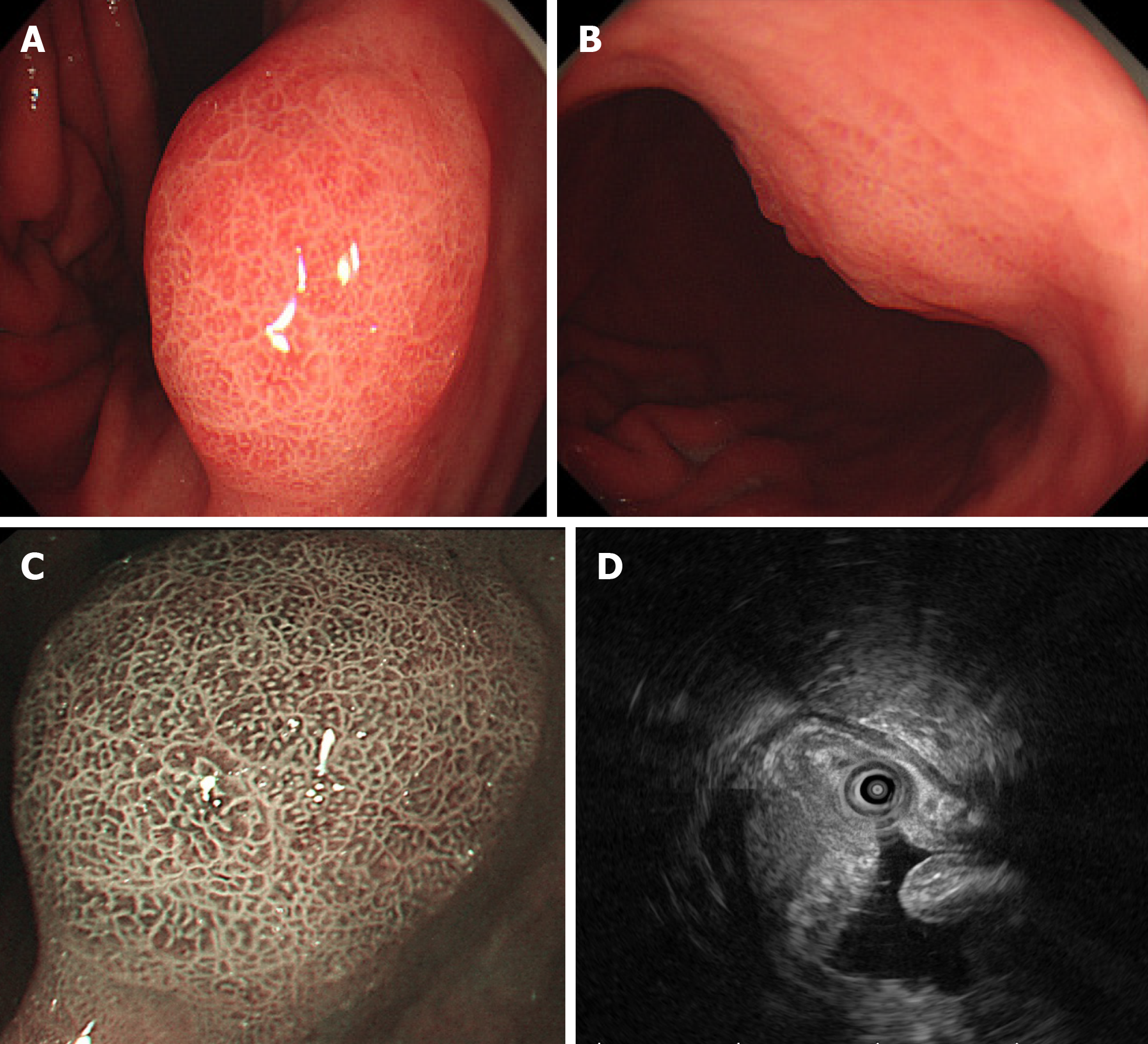
Figure 1 Endoscopic findings.
A and B: A 1.6-cm type 0-Is lesion covered by congested mucosa on gastric angle was observed by white light endoscopy; C: Regular microvascular pattern, presence of a demarcation line, irregular and smaller crypt opening, and widened intervening part were observed by magnifying endoscopy with narrow-band imaging. Magnification: 40×; D: Endoscopic ultrasound showed a 15.3 mm × 9 mm hypoechoic mass within the submucosal layer and thickened mucosal and submucosal layers.
- Citation: Cheng XL, Liu H. Gastric adenocarcinoma mimicking a submucosal tumor: A case report. World J Clin Cases 2019; 7(19): 3138-3144
- URL: https://www.wjgnet.com/2307-8960/full/v7/i19/3138.htm
- DOI: https://dx.doi.org/10.12998/wjcc.v7.i19.3138


