Copyright
©The Author(s) 2019.
World J Clin Cases. Oct 6, 2019; 7(19): 3111-3119
Published online Oct 6, 2019. doi: 10.12998/wjcc.v7.i19.3111
Published online Oct 6, 2019. doi: 10.12998/wjcc.v7.i19.3111
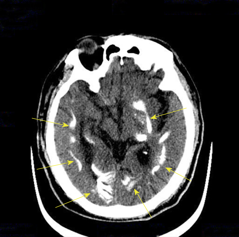
Figure 2 Transverse section of computed tomography scan.
The arrows point to multiple symmetrical patchy calcifications in the bilateral white matter, basal ganglia, thalamic area, and cerebellar hemisphere.
- Citation: Ding LN, Wang Y, Tian J, Ye LF, Chen S, Wu SM, Shang WB. Primary hypoparathyroidism accompanied by rhabdomyolysis induced by infection: A case report. World J Clin Cases 2019; 7(19): 3111-3119
- URL: https://www.wjgnet.com/2307-8960/full/v7/i19/3111.htm
- DOI: https://dx.doi.org/10.12998/wjcc.v7.i19.3111


