Copyright
©The Author(s) 2019.
World J Clin Cases. Sep 26, 2019; 7(18): 2910-2915
Published online Sep 26, 2019. doi: 10.12998/wjcc.v7.i18.2910
Published online Sep 26, 2019. doi: 10.12998/wjcc.v7.i18.2910
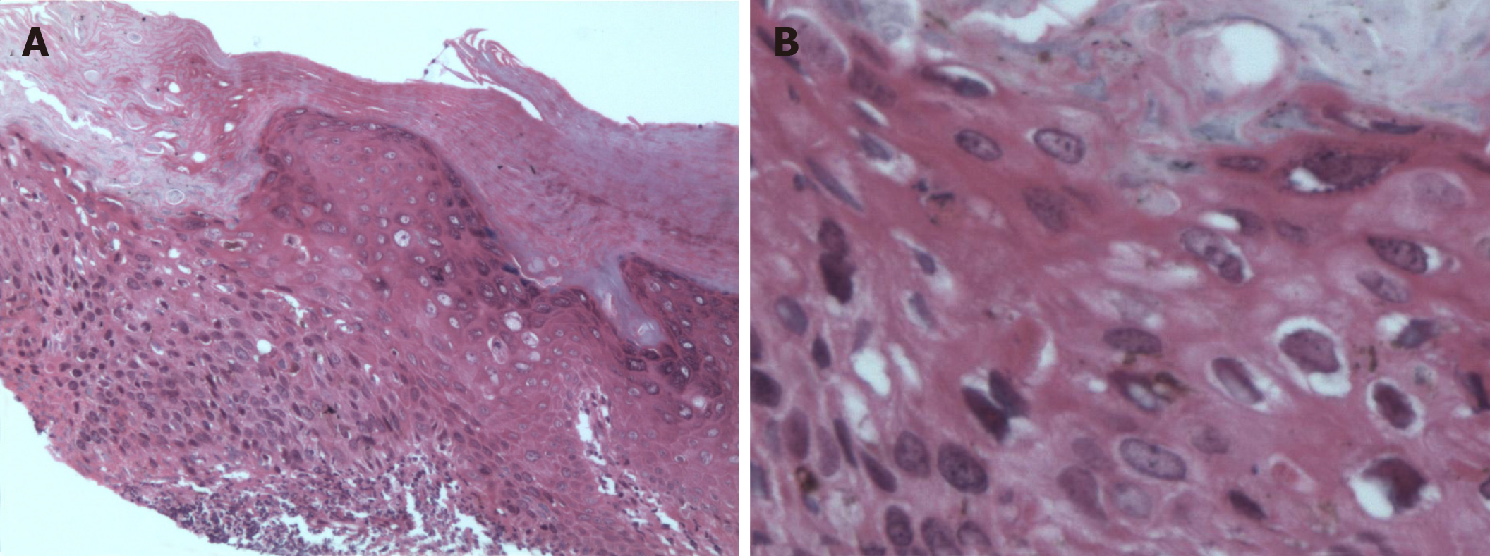
Figure 3 Pathohistological features of Bowen’s disease.
A and B: Tissue sections stained with hematoxylin and eosin (A: 100×; B: 200×). Histological characteristics in representative images include hyperkeratosis and parakeratosis, atypical keratinocytes throughout the epidermis, most mitotic phases, disordered cell arrangement, and dyskeratotic cells; additionally, some keratinocytes were vacuolated. A moderate amount of inflammatory infiltrate was present in the upper dermis, which is consistent with Bowen’s disease.
- Citation: Yu SR, Zhang JZ, Pu XM, Kang XJ. Bowen’s disease on the palm: A case report. World J Clin Cases 2019; 7(18): 2910-2915
- URL: https://www.wjgnet.com/2307-8960/full/v7/i18/2910.htm
- DOI: https://dx.doi.org/10.12998/wjcc.v7.i18.2910


