
















©The Author(s) 2019.
World J Clin Cases. Sep 26, 2019; 7(18): 2894-2898
Published online Sep 26, 2019. doi: 10.12998/wjcc.v7.i18.2894
Published online Sep 26, 2019. doi: 10.12998/wjcc.v7.i18.2894
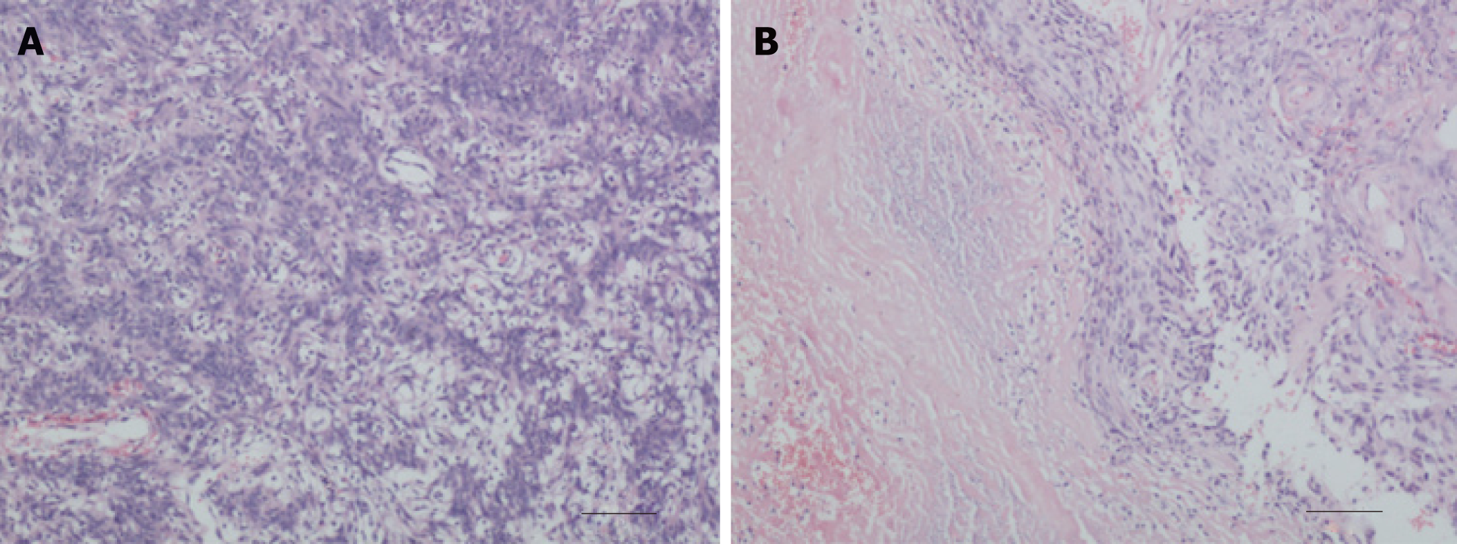
Figure 2 Entrapment of the temporal horn secondary to postoperative gamma-knife radiosurgery in intraventricular meningioma.
A: Pathology after first operation. Microscopically, the tumor cells were spindle-shaped and bundle-shaped with slightly larger and darker nuclei, and the diagnosis was meningioma; B: Pathology after second operation. Microscopically, the tumor cells were patchy, densely arranged, and slightly atypical, with massive necrosis and hemorrhage locally. The diagnosis was atypical meningioma.
- Citation: Liu J, Long SR, Li GY. Entrapment of the temporal horn secondary to postoperative gamma-knife radiosurgery in intraventricular meningioma: A case report. World J Clin Cases 2019; 7(18): 2894-2898
- URL: https://www.wjgnet.com/2307-8960/full/v7/i18/2894.htm
- DOI: https://dx.doi.org/10.12998/wjcc.v7.i18.2894