
















©The Author(s) 2019.
World J Clin Cases. Sep 26, 2019; 7(18): 2857-2863
Published online Sep 26, 2019. doi: 10.12998/wjcc.v7.i18.2857
Published online Sep 26, 2019. doi: 10.12998/wjcc.v7.i18.2857
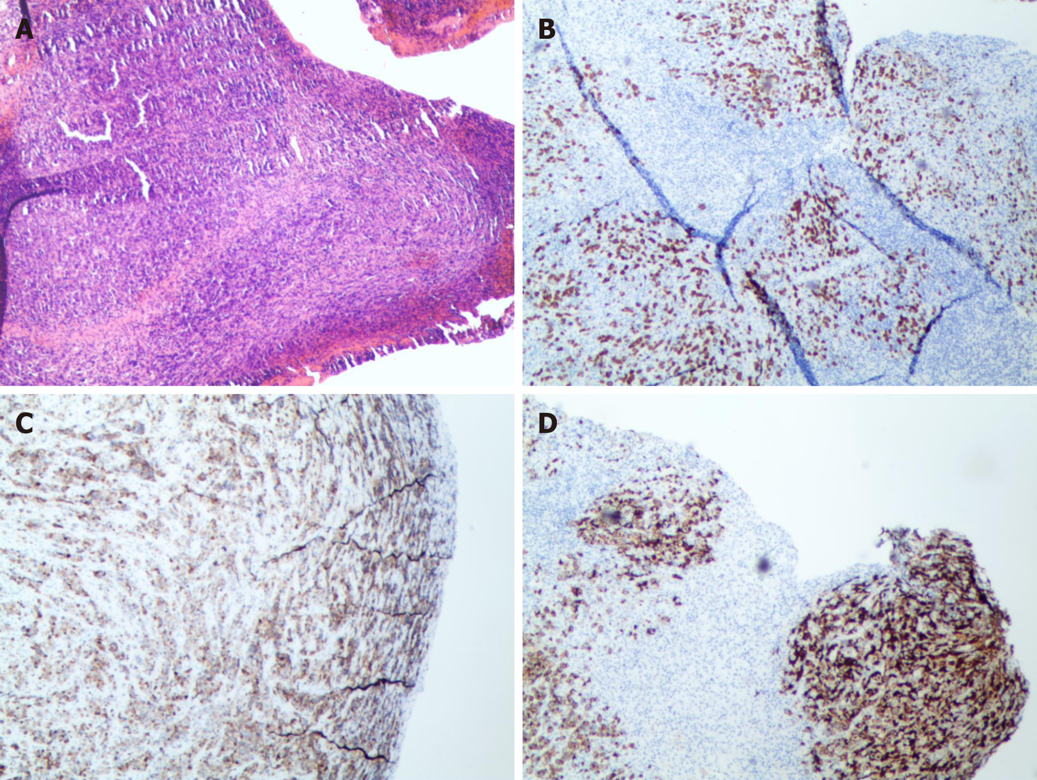
Figure 4 Low-magnification view of the hematoxylin and eosin staining shows diffuse infiltration of poorly differentiated mononuclear tumor cells (A).
Immunohistochemical staining (partial posting) showed positive results for CD3, CD30 and CD246 (ALK protein) (B–D).
- Citation: Yang S, Jiang WM, Yang HL. ALK-positive anaplastic large cell lymphoma of the thoracic spine occurring in pregnancy: A case report. World J Clin Cases 2019; 7(18): 2857-2863
- URL: https://www.wjgnet.com/2307-8960/full/v7/i18/2857.htm
- DOI: https://dx.doi.org/10.12998/wjcc.v7.i18.2857