Copyright
©The Author(s) 2019.
World J Clin Cases. Sep 26, 2019; 7(18): 2823-2830
Published online Sep 26, 2019. doi: 10.12998/wjcc.v7.i18.2823
Published online Sep 26, 2019. doi: 10.12998/wjcc.v7.i18.2823
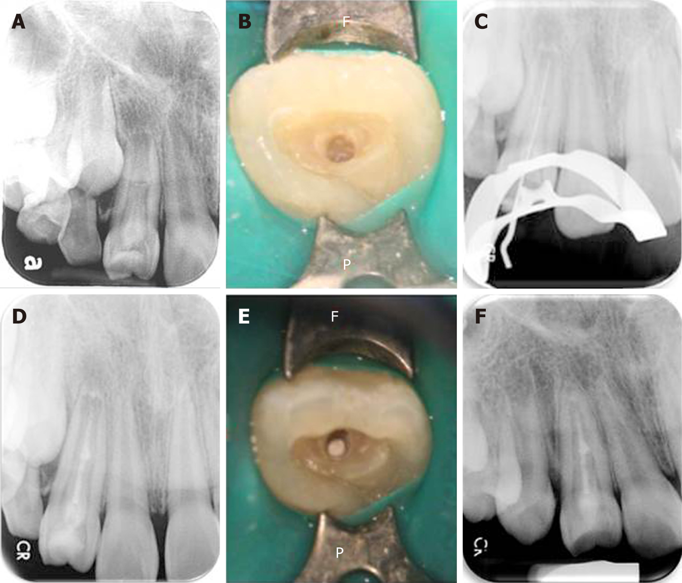
Figure 4 The operating process.
A: Pre-operative radiograph showing the presence of dens invaginatus associated with an open apex and a large periapical lesion. The middle part of the bulge was suspected to be connected to the main root canal; B: The position of the invagination was located under a microscope and access-opening was performed; C: A root ZX mini apex locator and a periapical radiograph were used to determine the working length before conducting complete debridement; D: Considering the interconnections between the root canals in the middle segment, a mineral trioxide aggregate (MTA) sealer was used to fill and seal the pseudo root canal and perform pulp capping in the main canal; E: The MTA filling as observed under the microscope; F: At the one-year follow-up, the apical lesion had healed well (the tooth wall had thickened and the apical foramen was closed). The tooth still responded normally to the pulp vitality test.
- Citation: Lee HN, Chen YK, Chen CH, Huang CY, Su YH, Huang YW, Chuang FH. Conservative pulp treatment for Oehlers type III dens invaginatus: A case report. World J Clin Cases 2019; 7(18): 2823-2830
- URL: https://www.wjgnet.com/2307-8960/full/v7/i18/2823.htm
- DOI: https://dx.doi.org/10.12998/wjcc.v7.i18.2823


