Copyright
©The Author(s) 2019.
World J Clin Cases. Sep 6, 2019; 7(17): 2587-2596
Published online Sep 6, 2019. doi: 10.12998/wjcc.v7.i17.2587
Published online Sep 6, 2019. doi: 10.12998/wjcc.v7.i17.2587
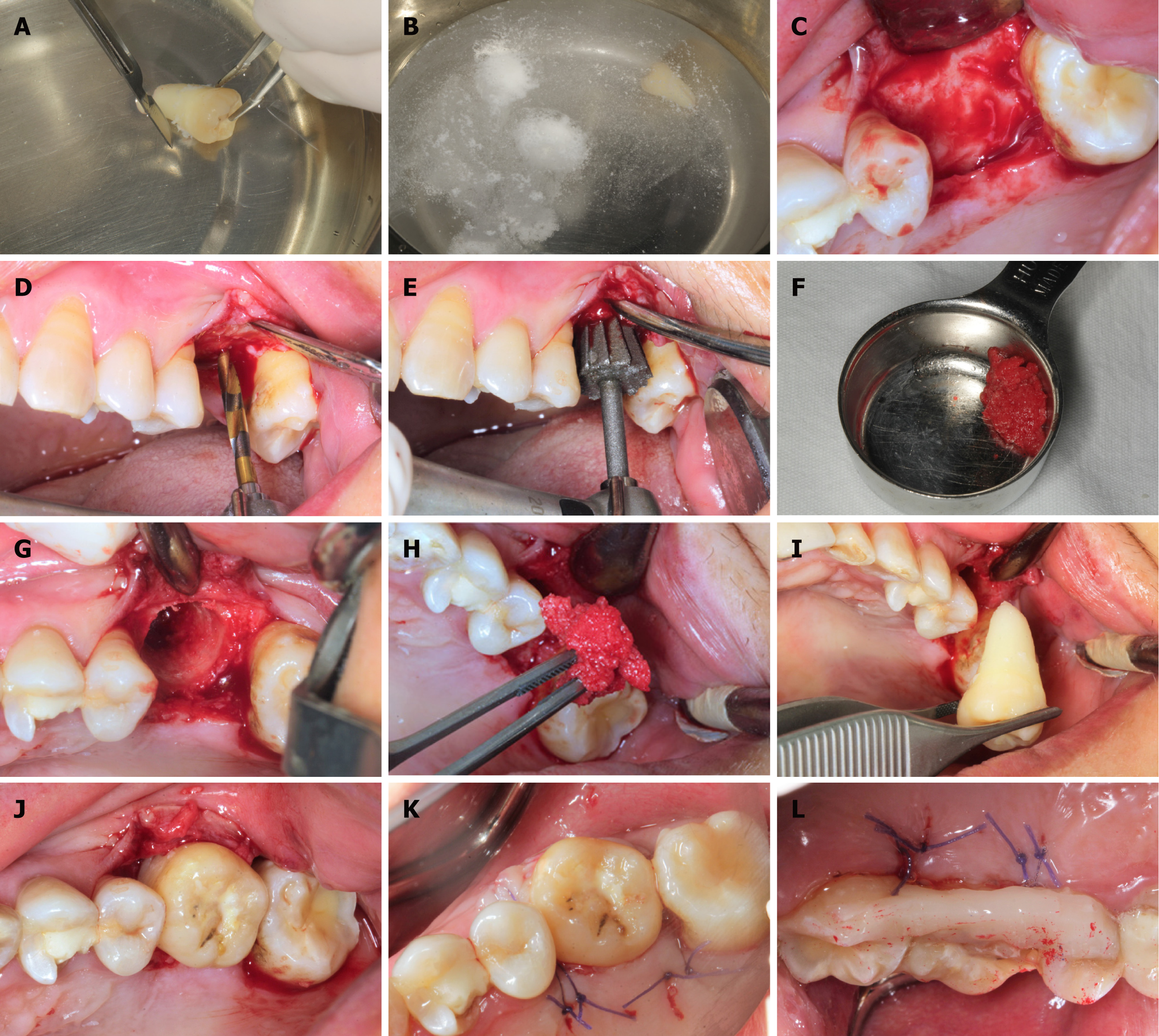
Figure 3 Surgical procedure.
A: The donor’s tooth was extracted, then the attached gingival was gently removed, but the periodontal ligament was kept; B: Treatment with solutions of 10 g/L cephalosporin, 75 g/L clindamycin hydrochloride, and 50 g/L aspirin in sequence; C: Soft tissue reflection; D: Expanding the pilot hole by using progressively wider drills; E: Finishing the preparation by using the individual drill; F: Collecting the autogenous bone during the hole preparation; G and H: Applying the autogenous bone to fill the compartment; I and J: Transplanting the tooth to the recipient’s site; K: Suture; L: Splinting the tooth with an acid-etch resin composite splint.
- Citation: Xu HD, Miron RJ, Zhang XX, Zhang YF. Allogenic tooth transplantation using 3D printing: A case report and review of the literature. World J Clin Cases 2019; 7(17): 2587-2596
- URL: https://www.wjgnet.com/2307-8960/full/v7/i17/2587.htm
- DOI: https://dx.doi.org/10.12998/wjcc.v7.i17.2587


