Copyright
©The Author(s) 2019.
World J Clin Cases. Sep 6, 2019; 7(17): 2587-2596
Published online Sep 6, 2019. doi: 10.12998/wjcc.v7.i17.2587
Published online Sep 6, 2019. doi: 10.12998/wjcc.v7.i17.2587
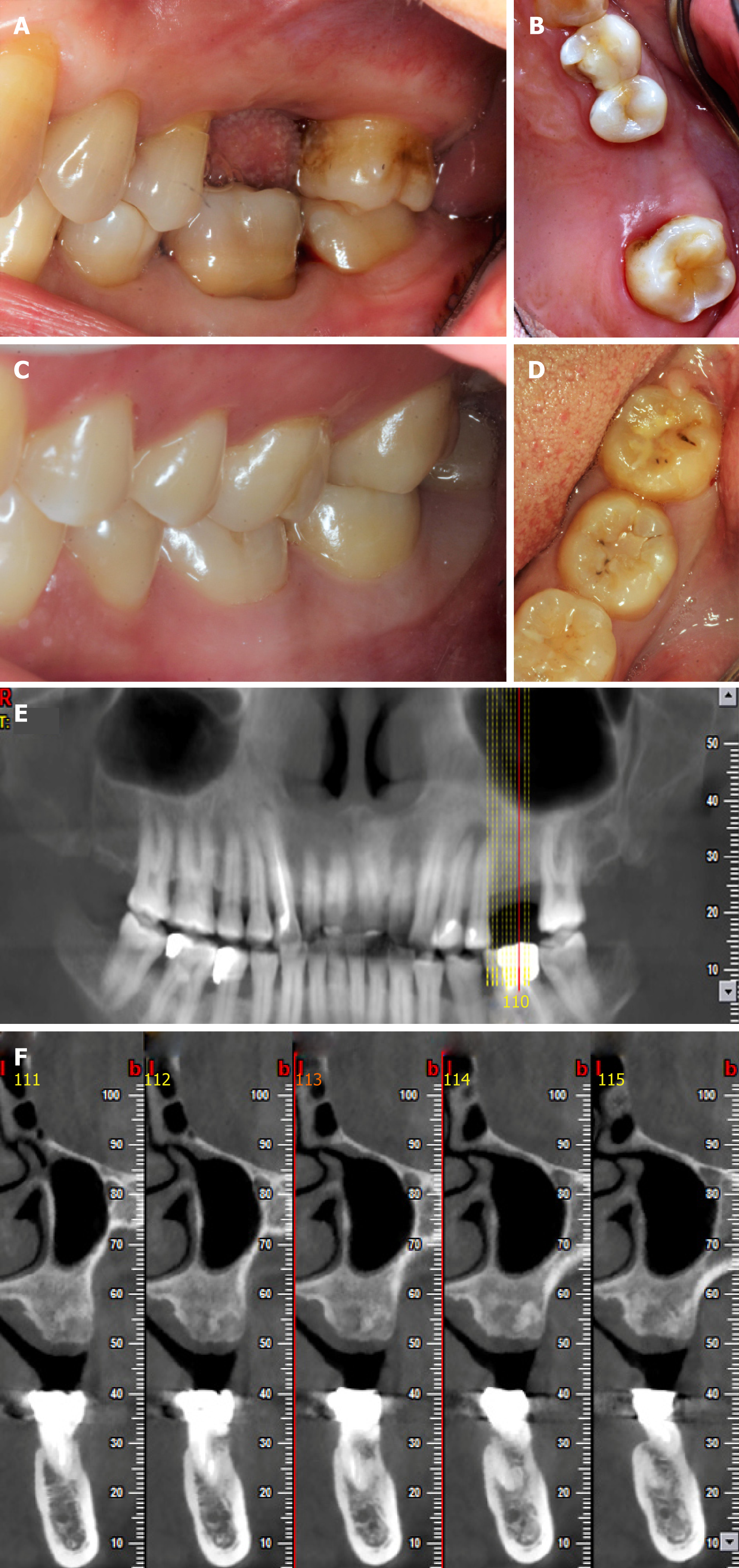
Figure 1 Preoperative photos and computed tomography scan of the donor and the recipient.
A and B: Occlusal photos showing the recipient’s missing #26; C and D: Occlusal photos showing the donor’s #48; E: The recipient’s orthopantomogram taken before the surgery; F: Computed tomography image showing the cross section of the bone at #26.
- Citation: Xu HD, Miron RJ, Zhang XX, Zhang YF. Allogenic tooth transplantation using 3D printing: A case report and review of the literature. World J Clin Cases 2019; 7(17): 2587-2596
- URL: https://www.wjgnet.com/2307-8960/full/v7/i17/2587.htm
- DOI: https://dx.doi.org/10.12998/wjcc.v7.i17.2587


