Copyright
©The Author(s) 2019.
World J Clin Cases. Aug 6, 2019; 7(15): 2094-2102
Published online Aug 6, 2019. doi: 10.12998/wjcc.v7.i15.2094
Published online Aug 6, 2019. doi: 10.12998/wjcc.v7.i15.2094
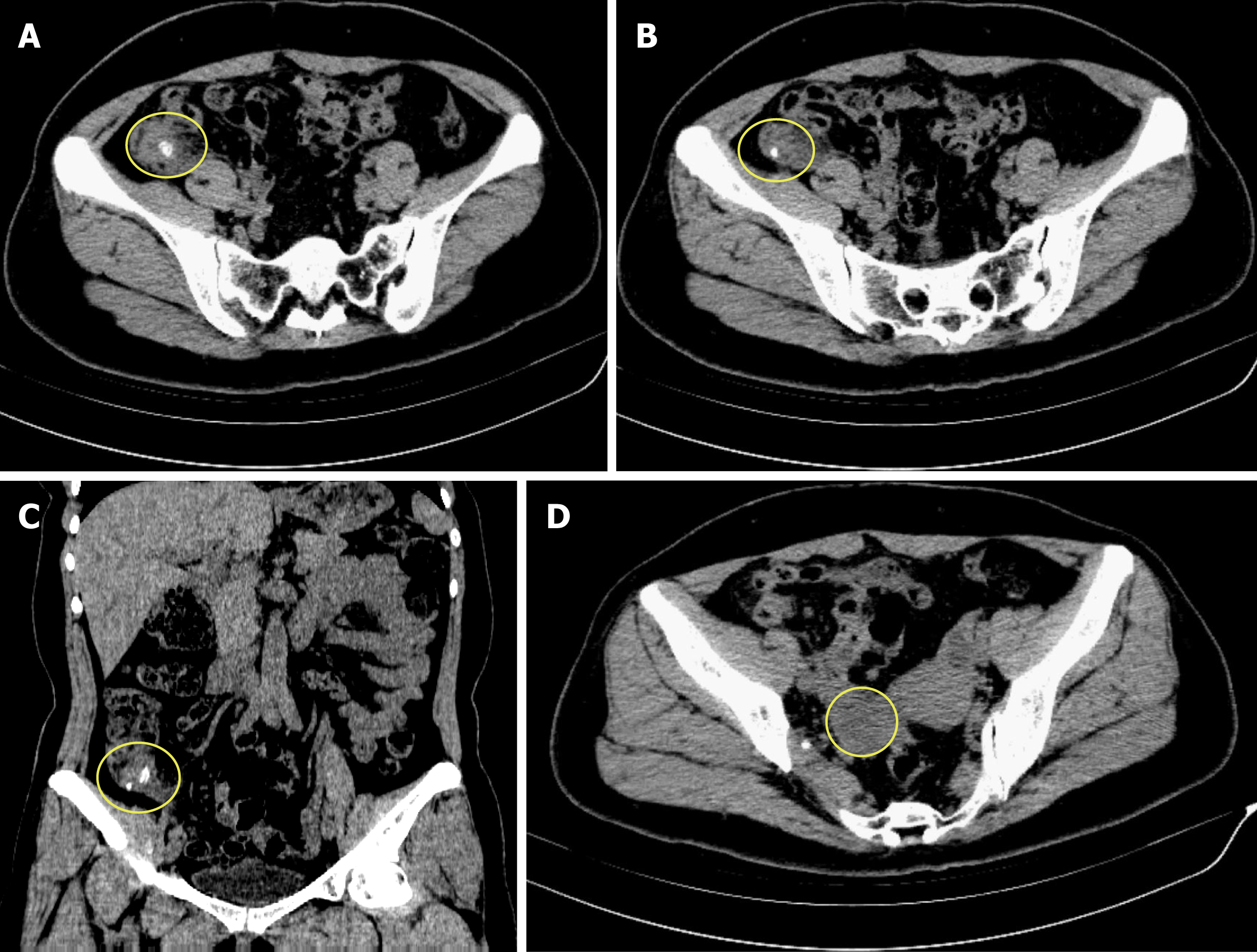
Figure 1 Computed tomography scan of the patient’s abdomen.
A and B: Transverse sections showing swelling of the appendix with surrounding exudation, and there is a fecalith at the root of the appendix, respectively; C: Coronal section showing swelling of the appendix with exudation and two separate fecaliths at its root; D: Transverse section showing a low density mass located in the right ovary.
- Citation: Zhu MY, Fei FM, Chen J, Zhou ZC, Wu B, Shen YY. Endometriosis of the duplex appendix: A case report and review of the literature. World J Clin Cases 2019; 7(15): 2094-2102
- URL: https://www.wjgnet.com/2307-8960/full/v7/i15/2094.htm
- DOI: https://dx.doi.org/10.12998/wjcc.v7.i15.2094


