Copyright
©The Author(s) 2019.
World J Clin Cases. Aug 6, 2019; 7(15): 2081-2086
Published online Aug 6, 2019. doi: 10.12998/wjcc.v7.i15.2081
Published online Aug 6, 2019. doi: 10.12998/wjcc.v7.i15.2081
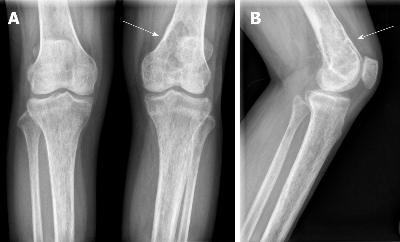
Figure 1 X-rays of knee joints.
A: Anteroposterior view; B: Lateral view. Bone trabeculae of both knees are sparse, bone density is widely reduced, and an oval osteolytic area is shown in the inferior medullary cavity of the left femur (arrow).
- Citation: Tang D, Wang XM, Zhang YS, Mi XX. Oncogenic osteomalacia caused by a phosphaturic mesenchymal tumor of the femur: A case report. World J Clin Cases 2019; 7(15): 2081-2086
- URL: https://www.wjgnet.com/2307-8960/full/v7/i15/2081.htm
- DOI: https://dx.doi.org/10.12998/wjcc.v7.i15.2081


