
















©The Author(s) 2019.
World J Clin Cases. Jul 26, 2019; 7(14): 1857-1864
Published online Jul 26, 2019. doi: 10.12998/wjcc.v7.i14.1857
Published online Jul 26, 2019. doi: 10.12998/wjcc.v7.i14.1857
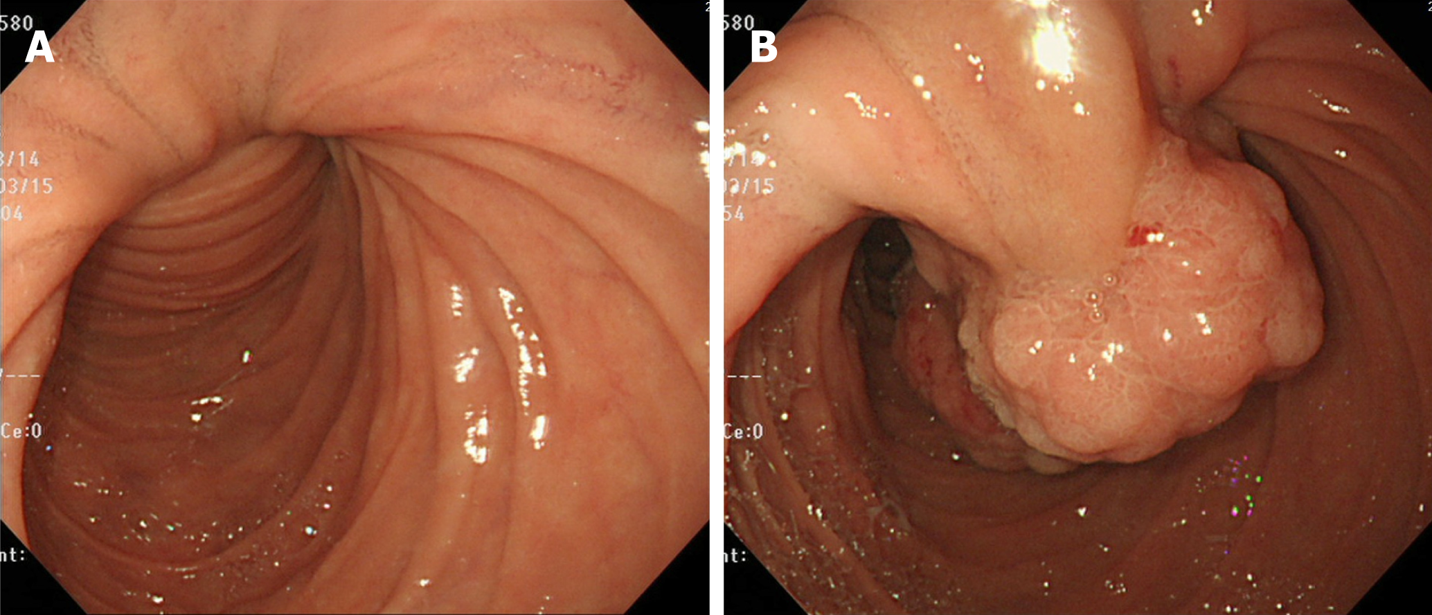
Figure 3 Esophagogastroduodenoscopy.
A: The mucosa of the duodenum is elongated distally; B: A pedunculate papillary polyp with minor bleeding located on the second part of the duodenum is observable when the scope is pushed. The ampulla is not identified.
- Citation: Hirata M, Shirakata Y, Yamanaka K. Duodenal intussusception secondary to ampullary adenoma: A case report. World J Clin Cases 2019; 7(14): 1857-1864
- URL: https://www.wjgnet.com/2307-8960/full/v7/i14/1857.htm
- DOI: https://dx.doi.org/10.12998/wjcc.v7.i14.1857