Copyright
©The Author(s) 2019.
World J Clin Cases. Jul 6, 2019; 7(13): 1726-1731
Published online Jul 6, 2019. doi: 10.12998/wjcc.v7.i13.1726
Published online Jul 6, 2019. doi: 10.12998/wjcc.v7.i13.1726
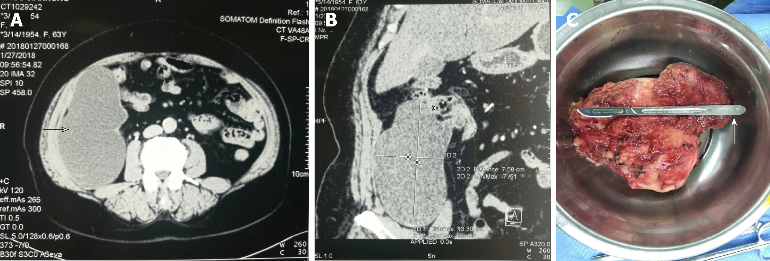
Figure 1 Computed tomography images.
A: Axial enhancement, cystic mass in the right iliac fossa, gourd-shaped, smooth margin, slight enhancement of cystic wall, slight delayed enhancement of mural nodules, and no enhancement of intracystic fluid (arrow); B. Coronal reconstruction showing that the lesion was located in the right lower abdominal ileocecal region and connected with the cecum wall (arrow); C: Pathological specimen showing the general view of the resected mass (at the arrow is the root of the appendix).
- Citation: Yang JM, Zhang WH, Yang DD, Jiang H, Yu L, Gao F. Giant low-grade appendiceal mucinous neoplasm: A case report. World J Clin Cases 2019; 7(13): 1726-1731
- URL: https://www.wjgnet.com/2307-8960/full/v7/i13/1726.htm
- DOI: https://dx.doi.org/10.12998/wjcc.v7.i13.1726


