Copyright
©The Author(s) 2019.
World J Clin Cases. Jul 6, 2019; 7(13): 1717-1725
Published online Jul 6, 2019. doi: 10.12998/wjcc.v7.i13.1717
Published online Jul 6, 2019. doi: 10.12998/wjcc.v7.i13.1717
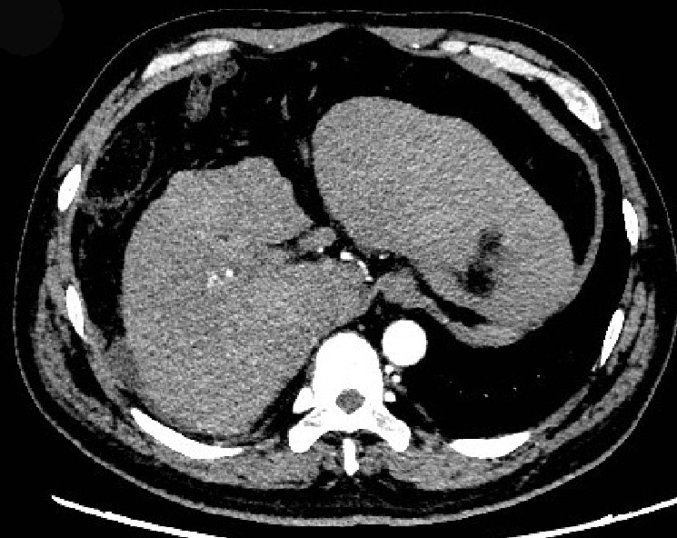
Figure 1 Contrast-enhanced computed tomography image.
The liver volume decreased. Computed tomography showed an irregular shape of the surface, the proportion of each leaf was not coordinated, and the density of the hepatic parenchymal was variable.
- Citation: Zhang MY, Niu JQ, Wen XY, Jin QL. Liver failure associated with benzbromarone: A case report and review of the literature. World J Clin Cases 2019; 7(13): 1717-1725
- URL: https://www.wjgnet.com/2307-8960/full/v7/i13/1717.htm
- DOI: https://dx.doi.org/10.12998/wjcc.v7.i13.1717


