Copyright
©The Author(s) 2019.
World J Clin Cases. Jun 26, 2019; 7(12): 1515-1521
Published online Jun 26, 2019. doi: 10.12998/wjcc.v7.i12.1515
Published online Jun 26, 2019. doi: 10.12998/wjcc.v7.i12.1515
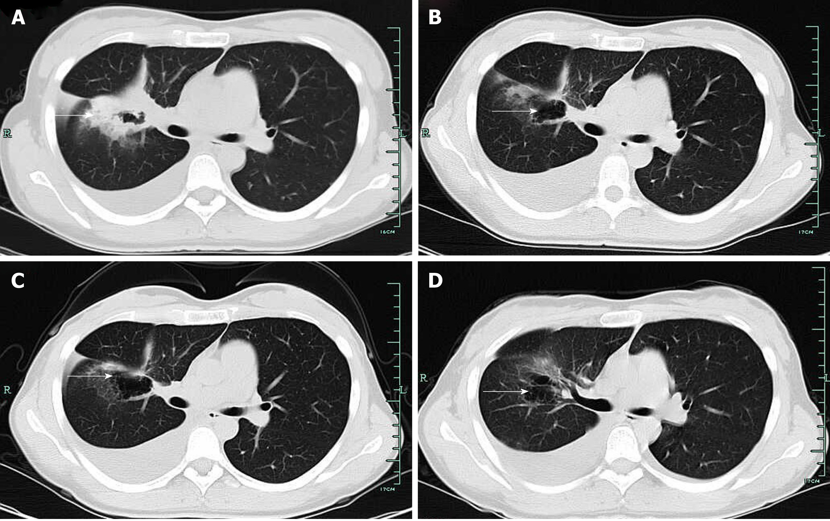
Figure 3 Case 2.
A: Computed tomography (CT) scan showed lung lesions with cavity, as well as pleural effusion before tyrosine kinase inhibitor treatment; B: CT showed a right pulmonary mass after 3 mo of ecotinib therapy, which resulted in a partial response after 3 mo; C: CT scan showed pleural effusion after 1 mo of bevacizumab (Avastin) and icotinib therapy; D: CT scan showed pleural effusion after 4 mo of bevacizumab (Avastin) and icotinib therapy, without progression of initial tumor.
- Citation: Yan RL, Wang J, Zhou JY, Chen Z, Zhou JY. Female genital tract metastasis of lung adenocarcinoma with EGFR mutations: Report of two cases. World J Clin Cases 2019; 7(12): 1515-1521
- URL: https://www.wjgnet.com/2307-8960/full/v7/i12/1515.htm
- DOI: https://dx.doi.org/10.12998/wjcc.v7.i12.1515


