Copyright
©The Author(s) 2019.
World J Clin Cases. Jun 26, 2019; 7(12): 1499-1507
Published online Jun 26, 2019. doi: 10.12998/wjcc.v7.i12.1499
Published online Jun 26, 2019. doi: 10.12998/wjcc.v7.i12.1499
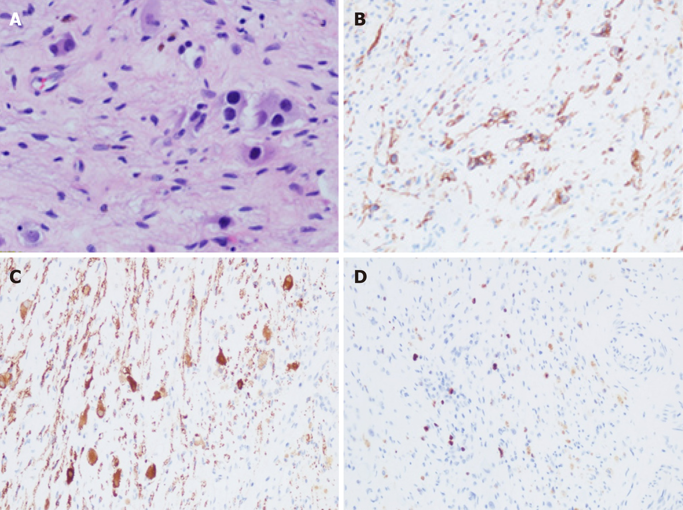
Figure 4 Pathological images.
A: Hematoxylin and eosin staining (40×); B-D: Immunohistochemical staining for CD56 (B), NSE (C), and Ki-67 (D) (20×).
- Citation: Chen DX, Hou YH, Jiang YN, Shao LW, Wang SJ, Wang XQ. Removal of pediatric stage IV neuroblastoma by robot-assisted laparoscopy: A case report and literature review. World J Clin Cases 2019; 7(12): 1499-1507
- URL: https://www.wjgnet.com/2307-8960/full/v7/i12/1499.htm
- DOI: https://dx.doi.org/10.12998/wjcc.v7.i12.1499


