Copyright
©The Author(s) 2019.
World J Clin Cases. Jun 26, 2019; 7(12): 1499-1507
Published online Jun 26, 2019. doi: 10.12998/wjcc.v7.i12.1499
Published online Jun 26, 2019. doi: 10.12998/wjcc.v7.i12.1499
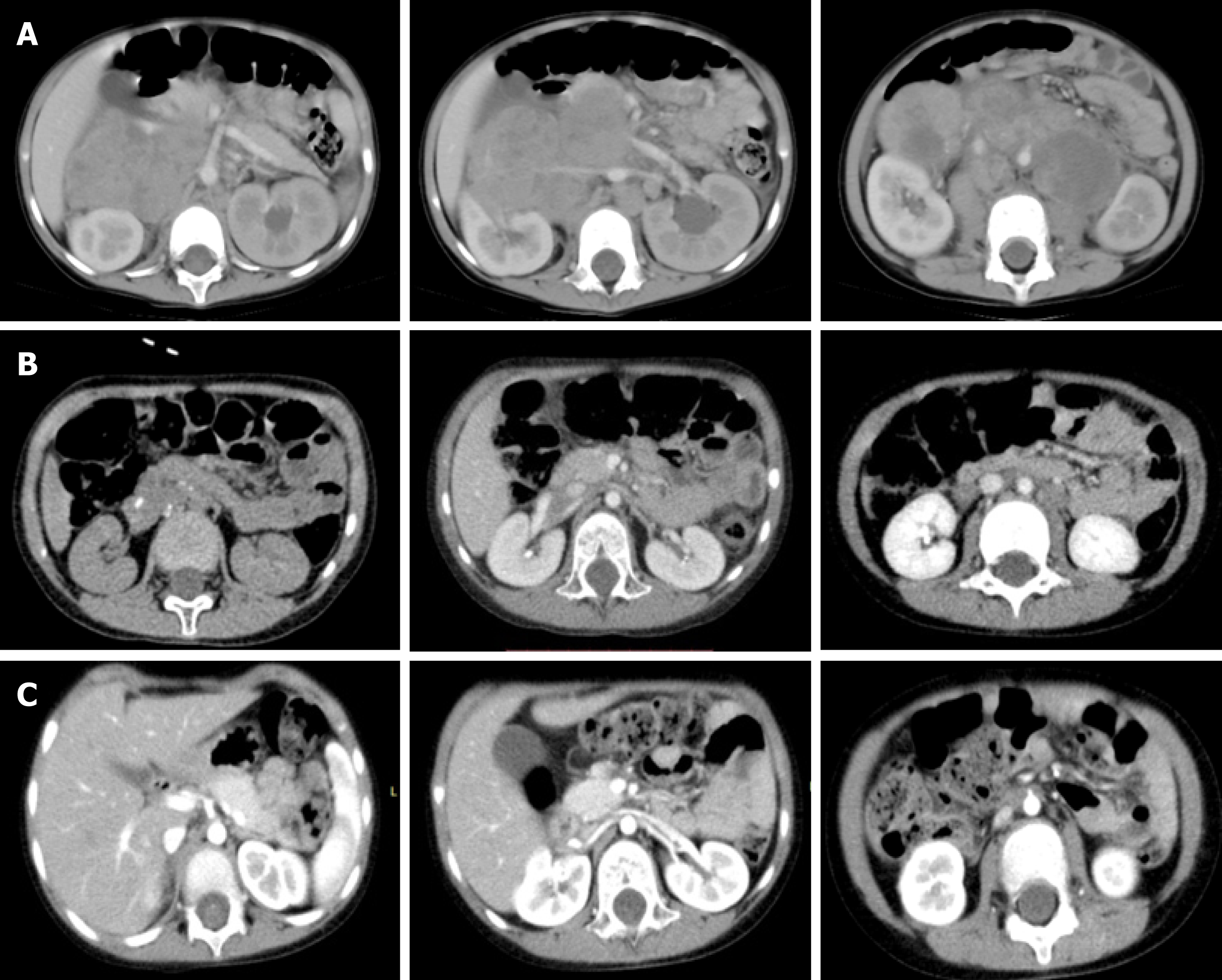
Figure 1 Abdominal enhanced computed tomography images.
A: A middle line retroperitoneal neoplasm with the inferior vena cava, right renal vessels, and abdominal aorta traversing in it (before chemotherapy); B: The tumor shrank considerably, but the above-mentioned vessels were still encased by the tumor (preoperatively after chemotherapy); C: Clearance of the tumor around the vessels (postoperative changes).
- Citation: Chen DX, Hou YH, Jiang YN, Shao LW, Wang SJ, Wang XQ. Removal of pediatric stage IV neuroblastoma by robot-assisted laparoscopy: A case report and literature review. World J Clin Cases 2019; 7(12): 1499-1507
- URL: https://www.wjgnet.com/2307-8960/full/v7/i12/1499.htm
- DOI: https://dx.doi.org/10.12998/wjcc.v7.i12.1499


