Copyright
©The Author(s) 2019.
World J Clin Cases. Jun 26, 2019; 7(12): 1410-1420
Published online Jun 26, 2019. doi: 10.12998/wjcc.v7.i12.1410
Published online Jun 26, 2019. doi: 10.12998/wjcc.v7.i12.1410
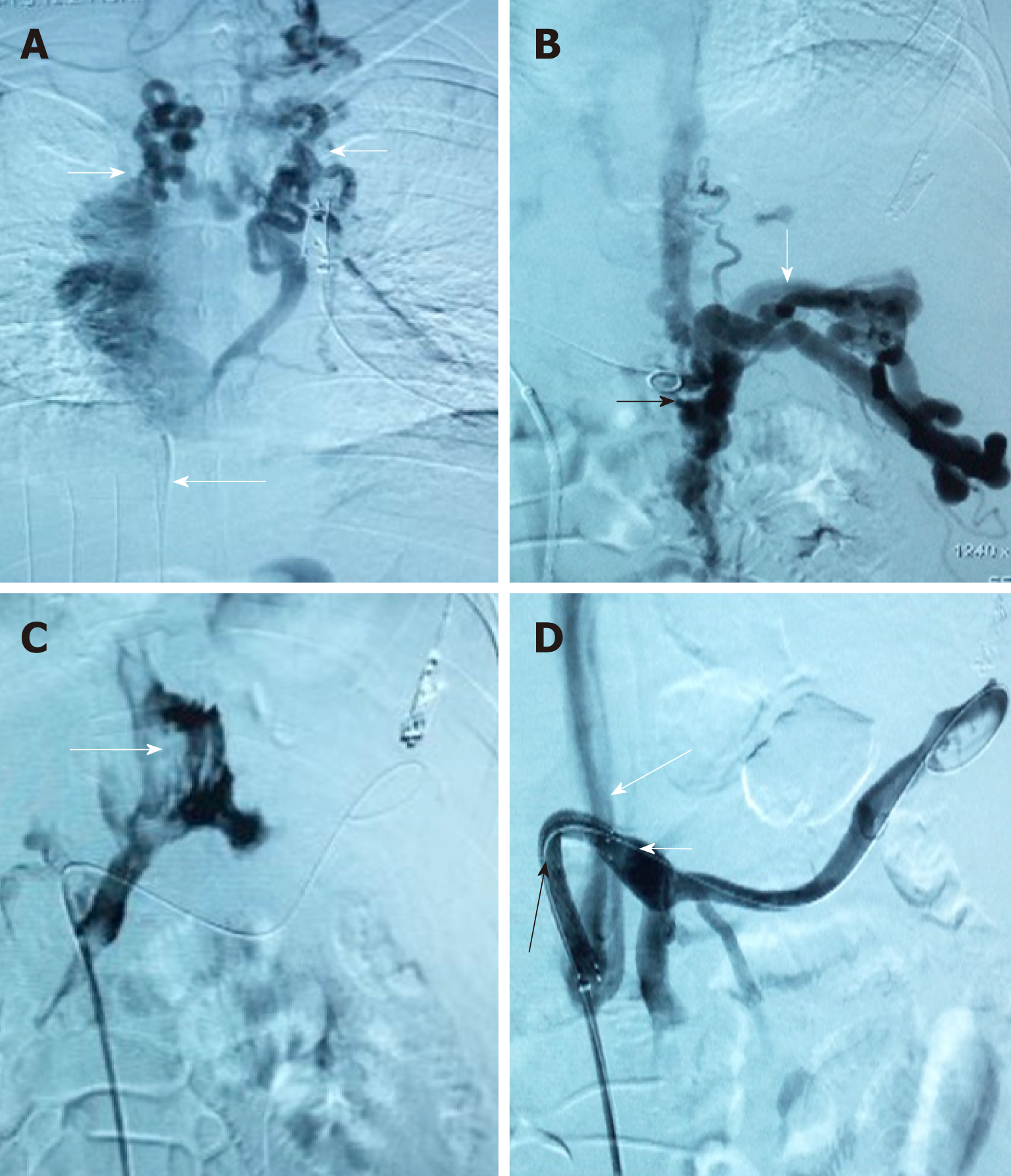
Figure 3 Due to complete occlusion of the bilateral jugular vein and/or superior vena cava, transjugular intrahepatic portosystemic shunt could not be performed in 3 cases.
A: Venogram via jugular vein access indicated that cavernous transformation of the bilateral jugular vein (white short arrow) and superior vena cava was occluded (long white arrow). Thus, conventional transjugular intrahepatic portosystemic shunt could not be performed; B: Puncture of the intrahepatic portal vein through the inferior vena cava via femoral vein access and venography. The venogram showed the angiography catheter in the portal vein (black arrow) and portal vein blood flow reflux into the hepatic varicose veins (long white arrow); C: The venogram showed that the contrast agent infiltrated the abdominal cavity (long white arrow) when the hepatic vein was punctured via the inferior vena cava; D: A semi-arc shunt between the portal vein (short white arrow) and the inferior vena cava (long white arrow) was successfully created using the covered stent (black arrow). Subsequently, the varicose veins disappeared completely.
- Citation: Zhang Y, Liu FQ, Yue ZD, Zhao HW, Wang L, Fan ZH, He FL. Safety and efficacy of transfemoral intrahepatic portosystemic shunt for portal hypertension: A single-center retrospective study. World J Clin Cases 2019; 7(12): 1410-1420
- URL: https://www.wjgnet.com/2307-8960/full/v7/i12/1410.htm
- DOI: https://dx.doi.org/10.12998/wjcc.v7.i12.1410


