
















©The Author(s) 2019.
World J Clin Cases. Jun 6, 2019; 7(11): 1337-1343
Published online Jun 6, 2019. doi: 10.12998/wjcc.v7.i11.1337
Published online Jun 6, 2019. doi: 10.12998/wjcc.v7.i11.1337
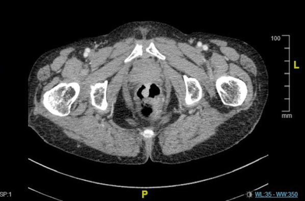
Figure 2 Enhanced pelvic computed tomography image revealing a pedunculated mass from the anterior wall of the rectum, and the other sessile mass from the side wall of the rectum.
Both masses invaded into muscular layer and were enhanced at the arterial phase.
- Citation: Cai YT, Cao LC, Zhu CF, Zhao F, Tian BX, Guo SY. Multiple synchronous anorectal melanomas with different colors: A case report. World J Clin Cases 2019; 7(11): 1337-1343
- URL: https://www.wjgnet.com/2307-8960/full/v7/i11/1337.htm
- DOI: https://dx.doi.org/10.12998/wjcc.v7.i11.1337