Copyright
©The Author(s) 2019.
World J Clin Cases. Jan 6, 2019; 7(1): 109-115
Published online Jan 6, 2019. doi: 10.12998/wjcc.v7.i1.109
Published online Jan 6, 2019. doi: 10.12998/wjcc.v7.i1.109
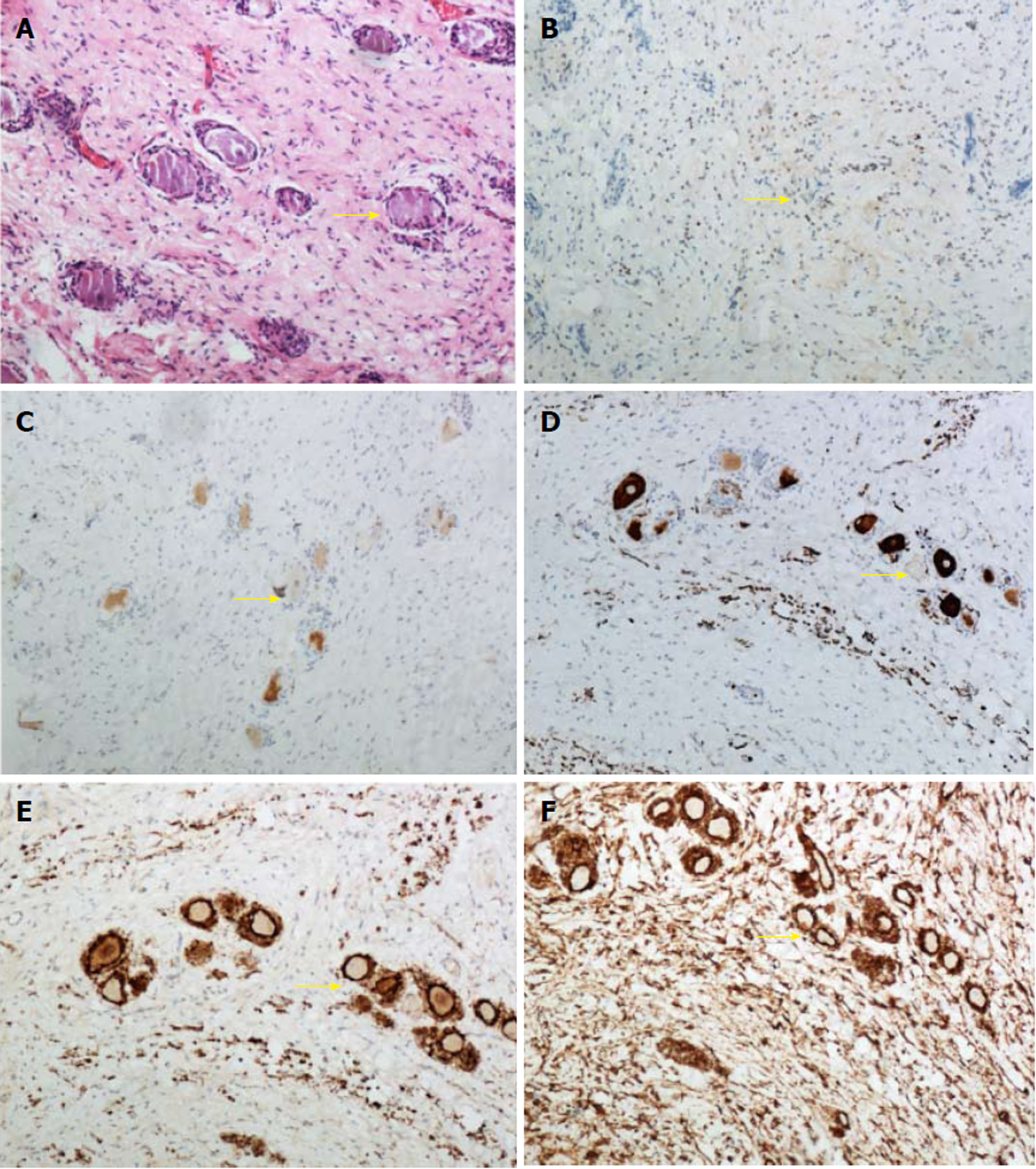
Figure 2 Histopathology of the resected lesion.
A: Hematoxylin and eosin staining showing spindle-shaped tumor cells, fusiform nuclei, and ganglion cells scattered in the tumor tissue (arrow). B-F: Immunohistochemistry showing Ki-67 (1%+) (B), NeuN (Scattered+) (C), NF (Scattered+) (D), S-100 (+) (E), and Vimentin (+) (F) (arrows).
- Citation: Tan CY, Liu JW, Lin Y, Tie XX, Cheng P, Qi X, Gao Y, Guo ZZ. Bilateral and symmetric C1-C2 dumbbell ganglioneuromas associated with neurofibromatosis type 1: A case report. World J Clin Cases 2019; 7(1): 109-115
- URL: https://www.wjgnet.com/2307-8960/full/v7/i1/109.htm
- DOI: https://dx.doi.org/10.12998/wjcc.v7.i1.109


