Copyright
©The Author(s) 2018.
World J Clin Cases. Sep 6, 2018; 6(9): 279-283
Published online Sep 6, 2018. doi: 10.12998/wjcc.v6.i9.279
Published online Sep 6, 2018. doi: 10.12998/wjcc.v6.i9.279
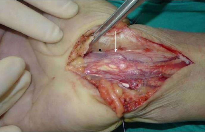
Figure 3 Photomicrograph of the surgical field.
After exposing the transverse carpal ligament, we identified the median nerve. The section of the median nerve coursing through the carpal canal had a significant bulge (white arrow) and a distal impression (black arrow).
- Citation: Luo PB, Zhang CQ. Chronic carpal tunnel syndrome caused by covert tophaceous gout: A case report. World J Clin Cases 2018; 6(9): 279-283
- URL: https://www.wjgnet.com/2307-8960/full/v6/i9/279.htm
- DOI: https://dx.doi.org/10.12998/wjcc.v6.i9.279


