Copyright
©The Author(s) 2018.
World J Clin Cases. Sep 6, 2018; 6(9): 279-283
Published online Sep 6, 2018. doi: 10.12998/wjcc.v6.i9.279
Published online Sep 6, 2018. doi: 10.12998/wjcc.v6.i9.279
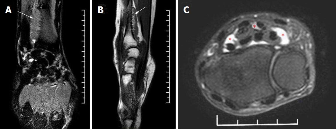
Figure 2 Magnetic resonance images of the patient’s right hand and wrist.
A: Lateral projection; B: Posteroanterior projection; C: Transverse projections. Magnetic resonance imaging revealed a giant mass adhering to the surface of the distal volar radius. The base of the mass was wide (A, B, white arrows). There was also a mass adjacent to the digital flexor tendons at the level of the proximal carpal tunnel. These were pressing on the median nerve (C, red asterisks).
- Citation: Luo PB, Zhang CQ. Chronic carpal tunnel syndrome caused by covert tophaceous gout: A case report. World J Clin Cases 2018; 6(9): 279-283
- URL: https://www.wjgnet.com/2307-8960/full/v6/i9/279.htm
- DOI: https://dx.doi.org/10.12998/wjcc.v6.i9.279


