Copyright
©The Author(s) 2018.
World J Clin Cases. Dec 26, 2018; 6(16): 1164-1168
Published online Dec 26, 2018. doi: 10.12998/wjcc.v6.i16.1164
Published online Dec 26, 2018. doi: 10.12998/wjcc.v6.i16.1164
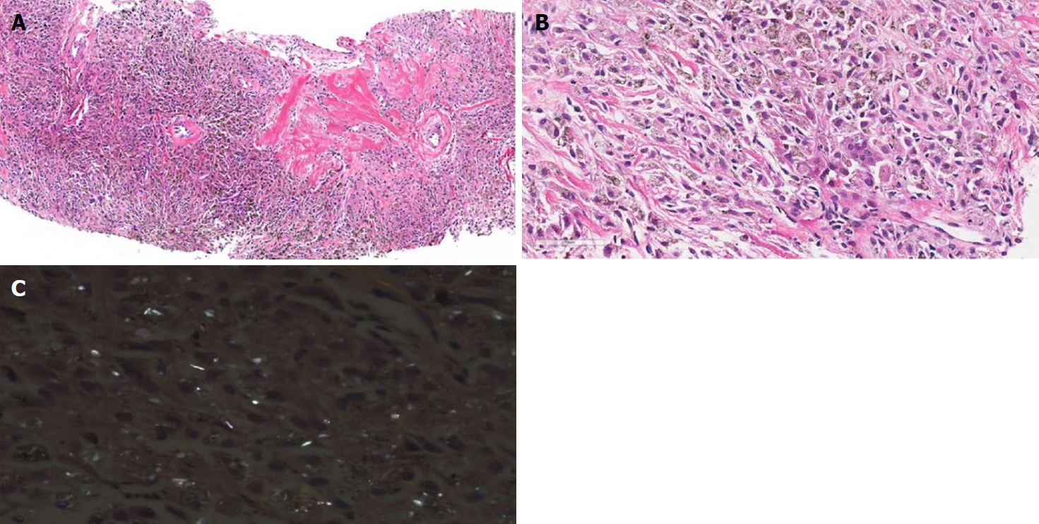
Figure 2 Histopathologic findings of lung biopsy specimen.
A: Lung parenchyma was replaced by diffuse infiltration of phagocytic macrophages with focal areas of sclerosis (original magnification, ×100); B: Macrophages contained anthracotic pigment and rare multinucleated giant cells (original magnification, ×400); C: Polarized microscopic examination revealed many birefringent crystals (original magnification, ×400).
- Citation: Yoon HY, Kim Y, Park HS, Kang CW, Ryu YJ. Combined silicosis and mixed dust pneumoconiosis with rapid progression: A case report and literature review. World J Clin Cases 2018; 6(16): 1164-1168
- URL: https://www.wjgnet.com/2307-8960/full/v6/i16/1164.htm
- DOI: https://dx.doi.org/10.12998/wjcc.v6.i16.1164


