Copyright
©The Author(s) 2018.
World J Clin Cases. Dec 6, 2018; 6(15): 1067-1072
Published online Dec 6, 2018. doi: 10.12998/wjcc.v6.i15.1067
Published online Dec 6, 2018. doi: 10.12998/wjcc.v6.i15.1067
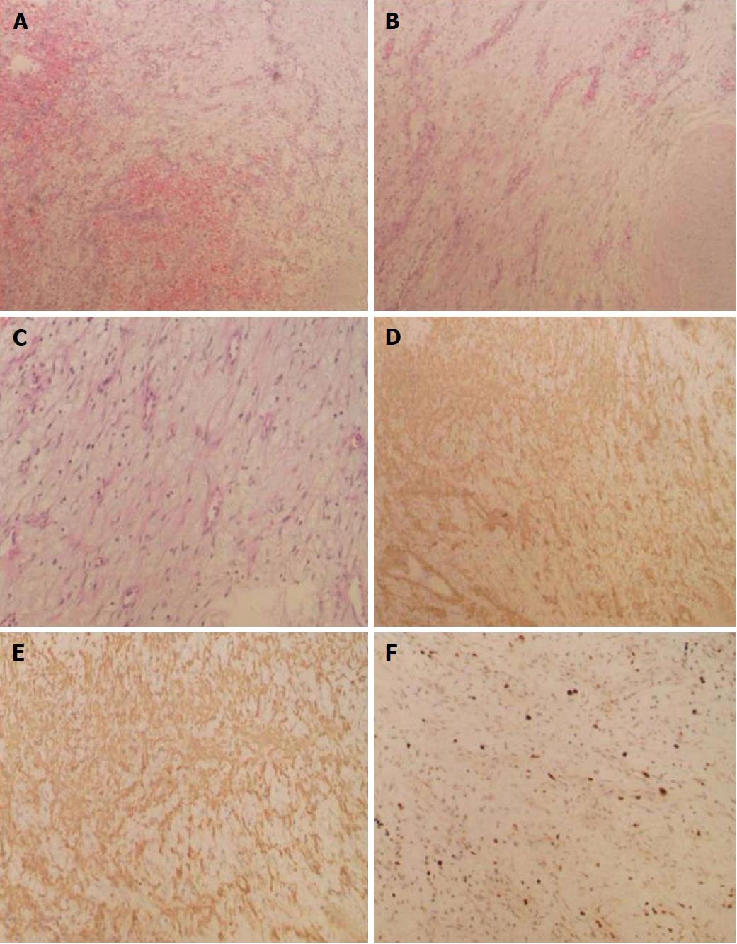
Figure 3 The diagnosis of small intestinal plexiform fibromyxoma.
A: Myxoid nodules and extensive hemorrhagic areas. The stroma was rich in small vessels (HE × 40); B: Tumor showed multinodular or plexiform growth, paucicellular nodules with blunt spindle cells and myxoid stroma (HE, × 40); C: Spindle-shaped bland tumor cells were separated by an abundant intercellular myxoid or fibromyxoid matrix (HE, × 100); D: Tumor SMA(+) (× 40); E: Tumor SMA(+) (× 100); F: Ki-67 labeling index < 5% (× 100). HE: Hematoxylin and eosin; SMA: Smooth muscle actin.
- Citation: Zhang WG, Xu LB, Xiang YN, Duan CH. Plexiform fibromyxoma of the small bowel: A case report. World J Clin Cases 2018; 6(15): 1067-1072
- URL: https://www.wjgnet.com/2307-8960/full/v6/i15/1067.htm
- DOI: https://dx.doi.org/10.12998/wjcc.v6.i15.1067


