
















©The Author(s) 2018.
World J Clin Cases. Nov 26, 2018; 6(14): 776-780
Published online Nov 26, 2018. doi: 10.12998/wjcc.v6.i14.776
Published online Nov 26, 2018. doi: 10.12998/wjcc.v6.i14.776
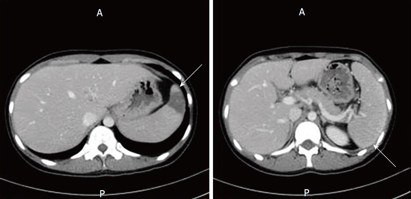
Figure 2 Thoracoabdominal contrast computed tomography scanning.
Computed tomography scan showing an area of low density in the spleen, suggesting splenic infarction (left arrow) and splenomegaly (right arrow).
- Citation: Miyaguchi K, Yamaoka M, Tsuzuki Y, Ashitani K, Ohgo H, Miyagawa Y, Ishizawa K, Kayano H, Nakamoto H, Imaeda H. Epstein–Barr virus-associated hemophagocytic syndrome in a patient with ulcerative colitis during treatment with azathioprine: A case report and review of literature. World J Clin Cases 2018; 6(14): 776-780
- URL: https://www.wjgnet.com/2307-8960/full/v6/i14/776.htm
- DOI: https://dx.doi.org/10.12998/wjcc.v6.i14.776