Copyright
©The Author(s) 2018.
World J Clin Cases. Nov 6, 2018; 6(13): 703-706
Published online Nov 6, 2018. doi: 10.12998/wjcc.v6.i13.703
Published online Nov 6, 2018. doi: 10.12998/wjcc.v6.i13.703
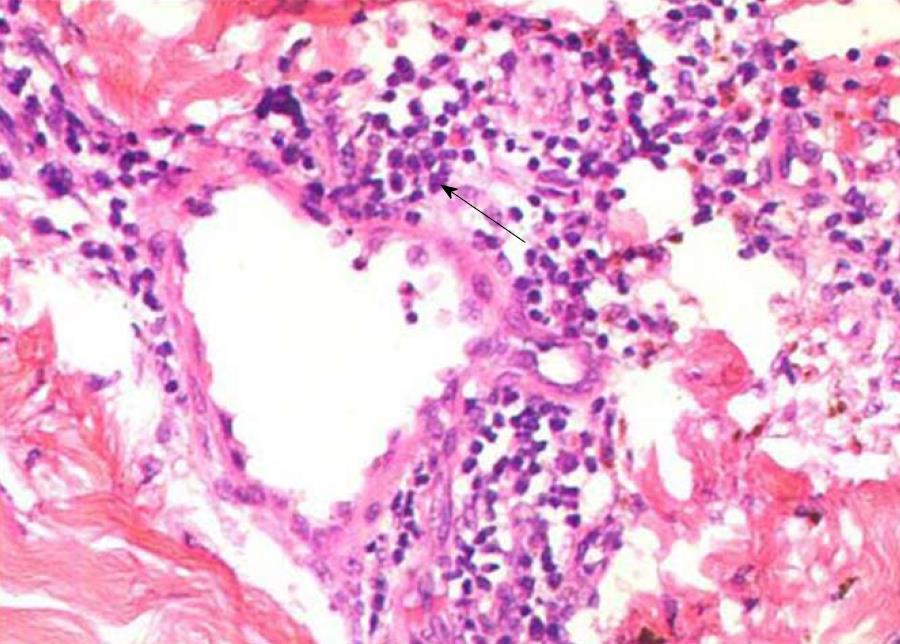
Figure 2 Light microscopy of the primary skin lesion.
Massive small lymphocytes (black arrow) are arranged around blood vessels throughout the dermis [hematoxylin and eosin (HE) staining, × 200]
- Citation: Li XL, Ma ZG, Huang WH, Chai EQ, Hao YF. Successful treatment of pyoderma gangrenosum with concomitant immunoglobulin A nephropathy: A case report and review of literature. World J Clin Cases 2018; 6(13): 703-706
- URL: https://www.wjgnet.com/2307-8960/full/v6/i13/703.htm
- DOI: https://dx.doi.org/10.12998/wjcc.v6.i13.703


