Copyright
©The Author(s) 2018.
World J Clin Cases. Oct 26, 2018; 6(12): 554-558
Published online Oct 26, 2018. doi: 10.12998/wjcc.v6.i12.554
Published online Oct 26, 2018. doi: 10.12998/wjcc.v6.i12.554
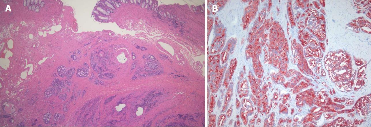
Figure 3 Histological images of metastatic prostate adenocarcinoma obtained from transanal full-thickness excisional biopsy.
A: Hematoxylin and eosin (HE) staining showed a moderate to poorly differentiated adenocarcinoma characterized by tumor cells diffusely infiltrated into submucosa and muscularis propria, sparing the mucosal layer (original magnification ×100); B: Immunohistochemical stains were performed and the tumor cells were positive for prostate specific antigen (PSA, original magnification ×100).
- Citation: You JH, Song JS, Jang KY, Lee MR. Computed tomography and magnetic resonance imaging findings of metastatic rectal linitis plastica from prostate cancer: A case report and review of literature. World J Clin Cases 2018; 6(12): 554-558
- URL: https://www.wjgnet.com/2307-8960/full/v6/i12/554.htm
- DOI: https://dx.doi.org/10.12998/wjcc.v6.i12.554


