Copyright
©The Author(s) 2018.
World J Clin Cases. Oct 6, 2018; 6(11): 459-465
Published online Oct 6, 2018. doi: 10.12998/wjcc.v6.i11.459
Published online Oct 6, 2018. doi: 10.12998/wjcc.v6.i11.459
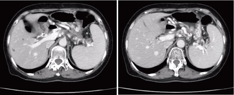
Figure 7 Follow-up contrast-enhanced computed tomography scan images.
Contrast-enhanced computed tomography of the abdomen demonstrating that the pseudocyst was completely resolved three months after stent removal. No reoccurrence was noted, as indicated by the significant resolution of the serious perisplenic and gastric varices.
- Citation: Wang BH, Xie LT, Zhao QY, Ying HJ, Jiang TA. Balloon dilator controls massive bleeding during endoscopic ultrasound-guided drainage for pancreatic pseudocyst: A case report and review of literature. World J Clin Cases 2018; 6(11): 459-465
- URL: https://www.wjgnet.com/2307-8960/full/v6/i11/459.htm
- DOI: https://dx.doi.org/10.12998/wjcc.v6.i11.459


