
















©The Author(s) 2018.
World J Clin Cases. Oct 6, 2018; 6(11): 459-465
Published online Oct 6, 2018. doi: 10.12998/wjcc.v6.i11.459
Published online Oct 6, 2018. doi: 10.12998/wjcc.v6.i11.459
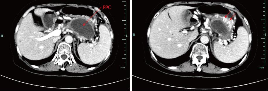
Figure 1 Pre-procedural contrast-enhanced computed tomography image.
Computed tomography (CT) scan of the upper abdomen images revealed a 7.4 cm × 6.2 cm pancreatic pseudocyst in the tail of the pancreas, which was in close contact with the posterior wall of the stomach. Notably, splenic vein occlusion, splenomegaly and gastric varices were also observed.
- Citation: Wang BH, Xie LT, Zhao QY, Ying HJ, Jiang TA. Balloon dilator controls massive bleeding during endoscopic ultrasound-guided drainage for pancreatic pseudocyst: A case report and review of literature. World J Clin Cases 2018; 6(11): 459-465
- URL: https://www.wjgnet.com/2307-8960/full/v6/i11/459.htm
- DOI: https://dx.doi.org/10.12998/wjcc.v6.i11.459