Copyright
©The Author(s) 2018.
World J Clin Cases. Sep 26, 2018; 6(10): 344-354
Published online Sep 26, 2018. doi: 10.12998/wjcc.v6.i10.344
Published online Sep 26, 2018. doi: 10.12998/wjcc.v6.i10.344
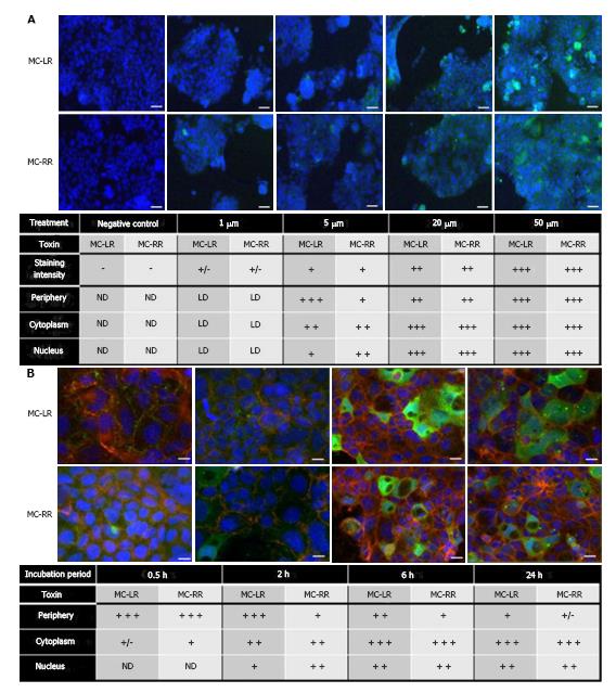
Figure 2 Subcellular localization of MC-LR and MC-RR in Caco-2 cells, which was adapted from a reference[11].
A: Caco-2 cells were treated for four hours with concentrations of MC-LR or MC-RR ranging from 1-50 μmol/L; B: Caco-2 cells were treated for several time points with 20 μmol/L of MC-LR or MC-RR. The cellular localization of these toxins was detected using an anti-ADDA antibody and an Alexa fluor 488 secondary antibody. Nuclei were counterstained with DAPI. The symbols (−), (+), (++) and (+++) represent the relative importance of the staining localization into the cell. Scale bars: A: 20 μm; B: 10 μm. ND: Alexa fluor 488 staining not detected; LD: Low signal of Alexa fluor 488 staining; (+/−) low signal intensity; MC: Microcystin.
- Citation: Wu JX, Huang H, Yang L, Zhang XF, Zhang SS, Liu HH, Wang YQ, Yuan L, Cheng XM, Zhuang DG, Zhang HZ. Gastrointestinal toxicity induced by microcystins. World J Clin Cases 2018; 6(10): 344-354
- URL: https://www.wjgnet.com/2307-8960/full/v6/i10/344.htm
- DOI: https://dx.doi.org/10.12998/wjcc.v6.i10.344


