Copyright
©The Author(s) 2018.
World J Clin Cases. Jan 16, 2018; 6(1): 6-10
Published online Jan 16, 2018. doi: 10.12998/wjcc.v6.i1.6
Published online Jan 16, 2018. doi: 10.12998/wjcc.v6.i1.6
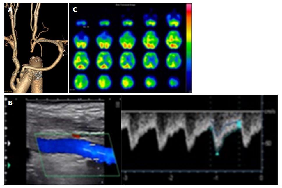
Figure 3 The findings of enhanced computed tomography, color duplex sonography and single-photon emission computed tomography after bypass.
A: Enhanced computed tomography findings. After the bypass procedure, the inflow is through the subclavian artery and the outflow is through the distal common carotid artery. Bifurcation of the external carotid artery and internal carotid artery is patent; B: Color duplex sonography findings. After bypass, the peak systolic velocity of the internal carotid artery was 60 cm/s; C: Single-photon emission computed tomography findings. After bypass, the cerebral blood flow was 38.39/31.04 mL/min per 100 g. The asymmetry ratio was 81%.
- Citation: Matsuda Y, Koyama T. Evaluation of revascularization after total arch replacement in common carotid artery occlusion. World J Clin Cases 2018; 6(1): 6-10
- URL: https://www.wjgnet.com/2307-8960/full/v6/i1/6.htm
- DOI: https://dx.doi.org/10.12998/wjcc.v6.i1.6


