Copyright
©The Author(s) 2017.
World J Clin Cases. Aug 16, 2017; 5(8): 333-339
Published online Aug 16, 2017. doi: 10.12998/wjcc.v5.i8.333
Published online Aug 16, 2017. doi: 10.12998/wjcc.v5.i8.333
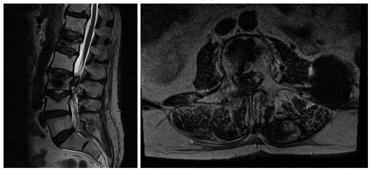
Figure 1 T2 weighed magnetic resonance imaging of the lumbar tract of the spinal column on sagittal and axial planes.
The images reveal the presence of a lesion located within the spinal channel at L2-L3. It is not possible to establish if it is located within the intradural or extradural space by the mere observation of the MRI. Note the needle trajectory inside the spinal channel at L1 on the right side.
- Citation: Tropeano MP, La Pira B, Pescatori L, Piccirilli M. Vertebroplasty and delayed subdural cauda equina hematoma: Review of literature and case report. World J Clin Cases 2017; 5(8): 333-339
- URL: https://www.wjgnet.com/2307-8960/full/v5/i8/333.htm
- DOI: https://dx.doi.org/10.12998/wjcc.v5.i8.333


