Copyright
©The Author(s) 2017.
World J Clin Cases. Jun 16, 2017; 5(6): 234-237
Published online Jun 16, 2017. doi: 10.12998/wjcc.v5.i6.234
Published online Jun 16, 2017. doi: 10.12998/wjcc.v5.i6.234
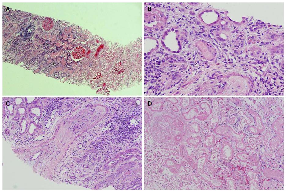
Figure 1 Renal pathology.
Pathological findings: A: Corticomedullary junction. The right side is necrotic renal cortex. On the left side of the image there is still viable corticomedullary junction. H and E, × 40; B: An arteriole obliterated with amorphous material (platelet and fibrin thrombus). H and E, × 200; C: An arcuate artery with almost completely obliterated lumen by severe fibrous to mucoid intimal thickening at the corticomedullary junction. H and E, × 100; D: Necrotic renal cortex without nuclear staining in the cells. H and E, × 200.
- Citation: Mansoor K, Zawodniak A, Nadasdy T, Khitan ZJ. Bilateral renal cortical necrosis associated with smoking synthetic cannabinoids. World J Clin Cases 2017; 5(6): 234-237
- URL: https://www.wjgnet.com/2307-8960/full/v5/i6/234.htm
- DOI: https://dx.doi.org/10.12998/wjcc.v5.i6.234


