Copyright
©The Author(s) 2017.
World J Clin Cases. Dec 16, 2017; 5(12): 440-445
Published online Dec 16, 2017. doi: 10.12998/wjcc.v5.i12.440
Published online Dec 16, 2017. doi: 10.12998/wjcc.v5.i12.440
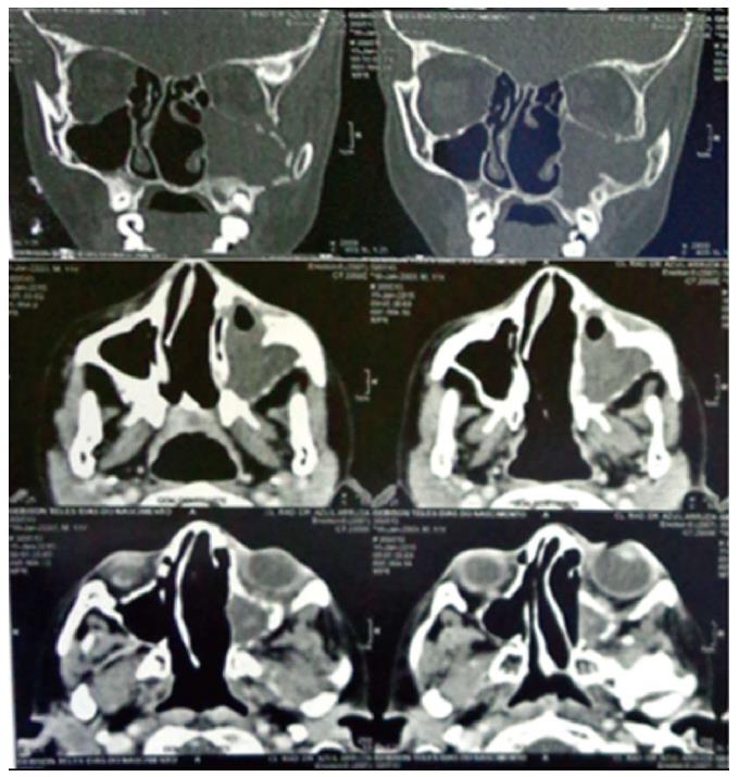
Figure 2 Computed tomography scan of the paranasal sinuses.
On coronal view, a diffuse hypodense mass was dislocating lateral wall of left sinus and compressing the inferior border of left orbital structure with tumor invasion. On axial plan, tumor mass was filling the left sinus and a dislocated nasal septum was evident.
- Citation: de Melo ACR, Lyra TC, Ribeiro ILA, da Paz AR, Bonan PRF, de Castro RD, Valença AMG. Embryonal rhabdomyosarcoma in the maxillary sinus with orbital involvement in a pediatric patient: Case report. World J Clin Cases 2017; 5(12): 440-445
- URL: https://www.wjgnet.com/2307-8960/full/v5/i12/440.htm
- DOI: https://dx.doi.org/10.12998/wjcc.v5.i12.440


