Copyright
©The Author(s) 2016.
World J Clin Cases. Jul 16, 2016; 4(7): 187-190
Published online Jul 16, 2016. doi: 10.12998/wjcc.v4.i7.187
Published online Jul 16, 2016. doi: 10.12998/wjcc.v4.i7.187
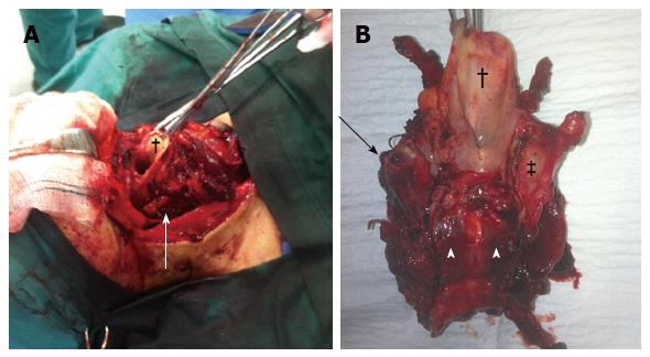
Figure 2 Intra-operative image showing the extent of the thyroid mass with absence of the thyroid cartilage (white arrow) (A) and posterior view of the excised larynx held by the clamps from the epiglottis (†) showed complete absence of the thyroid cartilage on the left (black arrow) some remnants of the thyroid cartilage on the right (‡) and posterior infiltration of the cricoid cartilage (arrow heads) (B).
- Citation: Georgiades F, Vasiliou G, Kyrodimos E, Thrasyvoulou G. Extensive laryngeal infiltration from a neglected papillary thyroid carcinoma: A case report. World J Clin Cases 2016; 4(7): 187-190
- URL: https://www.wjgnet.com/2307-8960/full/v4/i7/187.htm
- DOI: https://dx.doi.org/10.12998/wjcc.v4.i7.187


