Copyright
©The Author(s) 2016.
World J Clin Cases. Mar 16, 2016; 4(3): 94-98
Published online Mar 16, 2016. doi: 10.12998/wjcc.v4.i3.94
Published online Mar 16, 2016. doi: 10.12998/wjcc.v4.i3.94
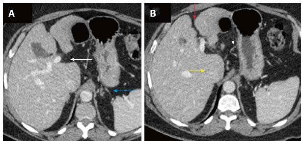
Figure 1 Triple phase dynamic contrast enhanced computed tomography performed for the upper abdomen showed features of chronic liver disease, predominantly on the portal venous phase.
A: Subtle sign of periportal space widening (white arrow) and few small collaterals in the peri-gastric location (blue arrow) suggesting features of chronic liver disease on computed tomography scan; B: Irregular liver margins seen better in the inter-lobar fissure (red arrow) with mild caudate enlargement (yellow arrow) and perigastric collaterals (white arrow).
- Citation: Laroia ST, Lata S. Hypertension in the liver clinic - polyarteritis nodosa in a patient with hepatitis B. World J Clin Cases 2016; 4(3): 94-98
- URL: https://www.wjgnet.com/2307-8960/full/v4/i3/94.htm
- DOI: https://dx.doi.org/10.12998/wjcc.v4.i3.94


