Copyright
©The Author(s) 2016.
World J Clin Cases. Mar 16, 2016; 4(3): 76-80
Published online Mar 16, 2016. doi: 10.12998/wjcc.v4.i3.76
Published online Mar 16, 2016. doi: 10.12998/wjcc.v4.i3.76
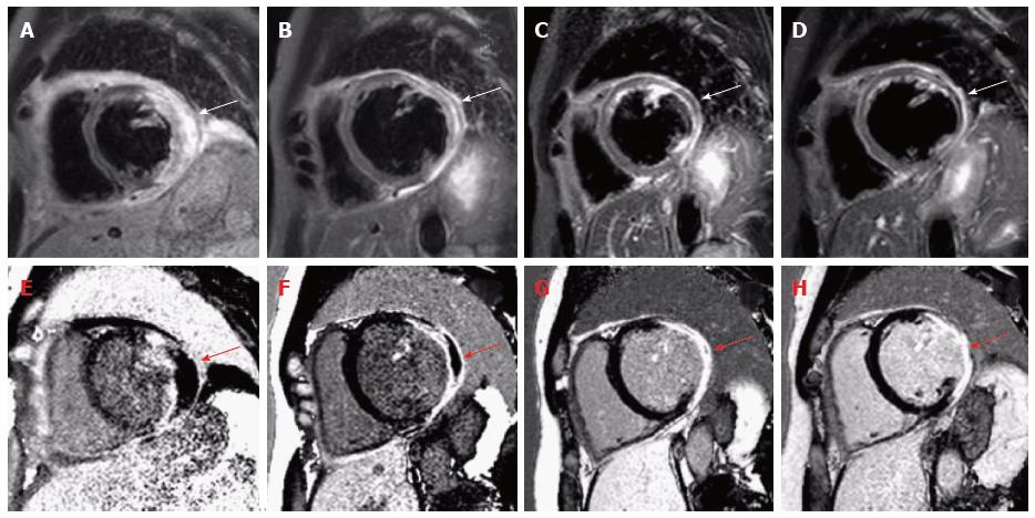
Figure 1 Note the hypointense core of the lesion on late gadolinium enhancement images, indicating massive myocardial injury with potentially myocardial hemorraghe, similar to the pattern of microvascular obstruction that can be found in patients with acute myocardial infarction.
T2-weighted short-tau inversion recovery images show hyperintense areas of myocardial edema (A) and necrosis/fibrosis on Late-Gadolinium-Enhancement images (E) of the left ventricular and septal wall before therapy (A + E). Cardiovascular magnetic resonance was repeated after 1 mo (B + F), 3 mo (C + G) and 6 mo (D + H) of glucocorticoid treatment demonstrating impressive resorption of edema and consolidation of scar.
- Citation: Fluschnik N, Lund G, Becher PM, Blankenberg S, Muellerleile K. Fulminant isolated cardiac sarcoidosis with pericardial effusion and acute heart failure: Challenging aspects of diagnosis and treatment. World J Clin Cases 2016; 4(3): 76-80
- URL: https://www.wjgnet.com/2307-8960/full/v4/i3/76.htm
- DOI: https://dx.doi.org/10.12998/wjcc.v4.i3.76


