Copyright
©The Author(s) 2016.
World J Clin Cases. Nov 16, 2016; 4(11): 364-368
Published online Nov 16, 2016. doi: 10.12998/wjcc.v4.i11.364
Published online Nov 16, 2016. doi: 10.12998/wjcc.v4.i11.364
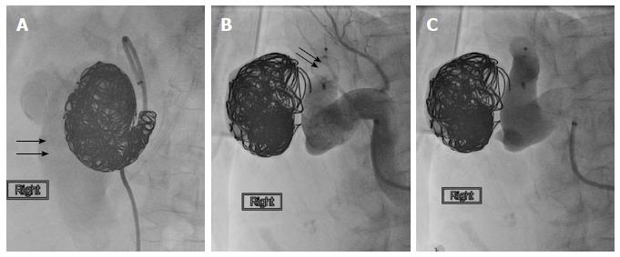
Figure 2 Conventional catheter angiogram image.
A: Conventional catheter angiogram image after using multiple coils showed persistent faint opacification of the inferior vena cava (arrows); B, C: Catheter angiogram image shows the placement of the Amplatzer vascular plug II (arrows) with successful obliteration of the fistulous communication. The proximal renal artery branches were spared.
- Citation: Nagpal P, Bathla G, Saboo SS, Khandelwal A, Goyal A, Rybicki FJ, Steigner ML. Giant idiopathic renal arteriovenous fistula managed by coils and amplatzer device: Case report and literature review. World J Clin Cases 2016; 4(11): 364-368
- URL: https://www.wjgnet.com/2307-8960/full/v4/i11/364.htm
- DOI: https://dx.doi.org/10.12998/wjcc.v4.i11.364


