Copyright
©The Author(s) 2015.
World J Clin Cases. Sep 16, 2015; 3(9): 838-842
Published online Sep 16, 2015. doi: 10.12998/wjcc.v3.i9.838
Published online Sep 16, 2015. doi: 10.12998/wjcc.v3.i9.838
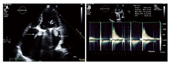
Figure 1 Prosthetic mitral valve thrombosis.
A: Transthoracic two-dimensional echocardiogram in apical four-chamber view; B: Transthoracic continuous wave Doppler across mitral valve. Bi-leaflet prosthetic mitral valve seen with echo density, suggestive of thrombus, attached to the atrial side of the bi-leaflet prosthetic mitral valve. The maximum peak gradient across the prosthetic mitral valve is 14 mmHg and mean peak gradient 4 mmHg.
- Citation: Mehra S, Movahed A, Espinoza C, Marcu CB. Horseshoe thrombus in a patient with mechanical prosthetic mitral valve: A case report and review of literature. World J Clin Cases 2015; 3(9): 838-842
- URL: https://www.wjgnet.com/2307-8960/full/v3/i9/838.htm
- DOI: https://dx.doi.org/10.12998/wjcc.v3.i9.838


