Copyright
©The Author(s) 2015.
World J Clin Cases. Sep 16, 2015; 3(9): 793-806
Published online Sep 16, 2015. doi: 10.12998/wjcc.v3.i9.793
Published online Sep 16, 2015. doi: 10.12998/wjcc.v3.i9.793
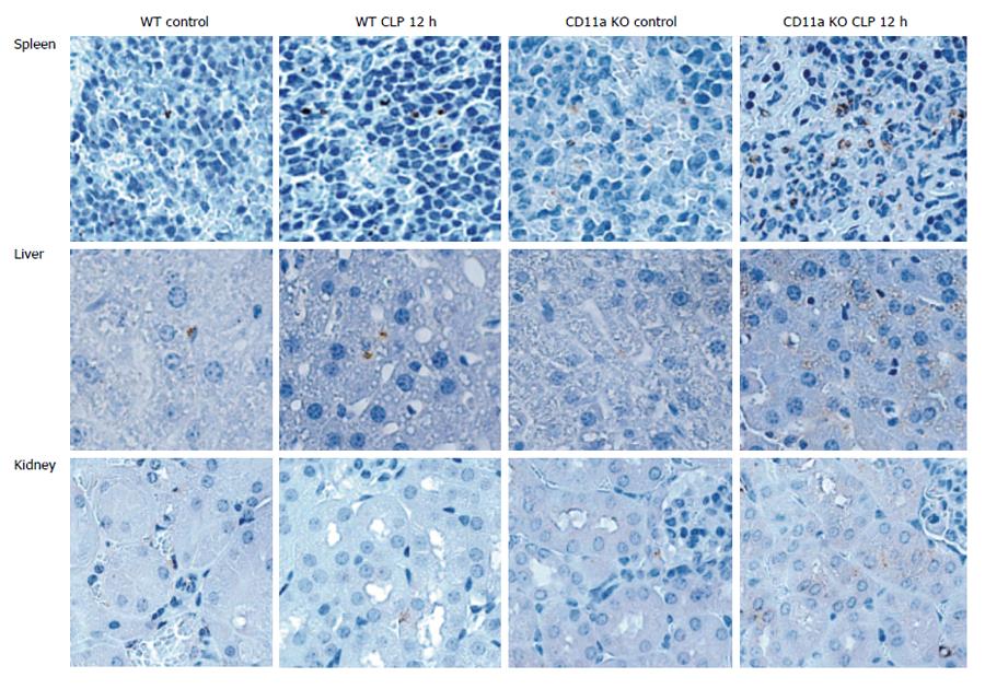
Figure 10 Neutrophil recruitment to tissues.
Neutrophil recruitment to tissues was studied by examining myeloperoxidase staining. Cells stained with brown show myeloperoxidase positive cells, largely representing neutrophils. In figures, control means no cecal ligation and puncture surgery, and cecal ligation and puncture (CLP) 12 h is 12 h after cecal ligatation and puncture model.
- Citation: Liu JR, Han X, Soriano SG, Yuki K. Leukocyte function-associated antigen-1 deficiency impairs responses to polymicrobial sepsis. World J Clin Cases 2015; 3(9): 793-806
- URL: https://www.wjgnet.com/2307-8960/full/v3/i9/793.htm
- DOI: https://dx.doi.org/10.12998/wjcc.v3.i9.793


