Copyright
©The Author(s) 2015.
World J Clin Cases. Sep 16, 2015; 3(9): 793-806
Published online Sep 16, 2015. doi: 10.12998/wjcc.v3.i9.793
Published online Sep 16, 2015. doi: 10.12998/wjcc.v3.i9.793
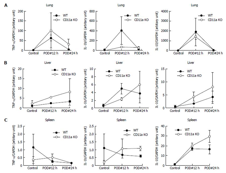
Figure 7 Tissues tumor necrosis factor-α, interleukin-1b and interleukin-10 levels.
Various tissues of IL-6 expression were examined using real-time quantitative polymerase chain reaction. Data represent mean ± SD of 4 independent experiments. Statistical analysis was performed using two-way analysis of variance with Tukey post hoc analysis. IL: Interleukin; TNF: Tumor necrosis factor; GAPDH: Glyceraldehyde-3-phosphate dehydrogenase.
- Citation: Liu JR, Han X, Soriano SG, Yuki K. Leukocyte function-associated antigen-1 deficiency impairs responses to polymicrobial sepsis. World J Clin Cases 2015; 3(9): 793-806
- URL: https://www.wjgnet.com/2307-8960/full/v3/i9/793.htm
- DOI: https://dx.doi.org/10.12998/wjcc.v3.i9.793


