Copyright
©The Author(s) 2015.
World J Clin Cases. Jul 16, 2015; 3(7): 661-670
Published online Jul 16, 2015. doi: 10.12998/wjcc.v3.i7.661
Published online Jul 16, 2015. doi: 10.12998/wjcc.v3.i7.661
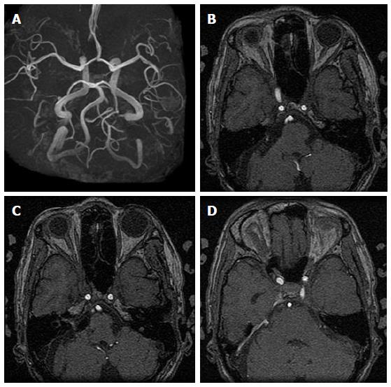
Figure 8 Postoperative three-dimensional time-of-flight magnetic resonance angiography (3D-TOF-MRA) and magnetic resonance angiographic source imaging showing anterior inferior cerebral artery (AICA) stump with disappearance of proximal and distal right AICA aneurysms (A, B), decreased intensity of right AICA and seemingly disappearance of proximal aneurysm, clip artifact at the entrance to the internal auditory canal and decreased intensity of intracanicular nidus (C), and persistence of prominent right superior petrosal sinus venous shunt (D).
- Citation: Tucker A, Tsuji M, Yamada Y, Hanabusa K, Ukita T, Miyake H, Ohmura T. Arteriovenous malformation of the vestibulocochlear nerve. World J Clin Cases 2015; 3(7): 661-670
- URL: https://www.wjgnet.com/2307-8960/full/v3/i7/661.htm
- DOI: https://dx.doi.org/10.12998/wjcc.v3.i7.661


