Copyright
©The Author(s) 2015.
World J Clin Cases. Jul 16, 2015; 3(7): 661-670
Published online Jul 16, 2015. doi: 10.12998/wjcc.v3.i7.661
Published online Jul 16, 2015. doi: 10.12998/wjcc.v3.i7.661
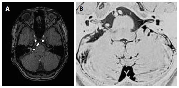
Figure 3 Magnetic resonance angiographic source imaging (A) and magnetic resonance cisternography (B) reveal a small (11.
4 mm × 4.2 mm) relatively compact nidus located in the cerebellopontine angle at the cisternal portion of the cranial nerves VII and VIII with extension into the internal auditory canal.
- Citation: Tucker A, Tsuji M, Yamada Y, Hanabusa K, Ukita T, Miyake H, Ohmura T. Arteriovenous malformation of the vestibulocochlear nerve. World J Clin Cases 2015; 3(7): 661-670
- URL: https://www.wjgnet.com/2307-8960/full/v3/i7/661.htm
- DOI: https://dx.doi.org/10.12998/wjcc.v3.i7.661


