Copyright
©The Author(s) 2015.
World J Clin Cases. Jul 16, 2015; 3(7): 607-613
Published online Jul 16, 2015. doi: 10.12998/wjcc.v3.i7.607
Published online Jul 16, 2015. doi: 10.12998/wjcc.v3.i7.607
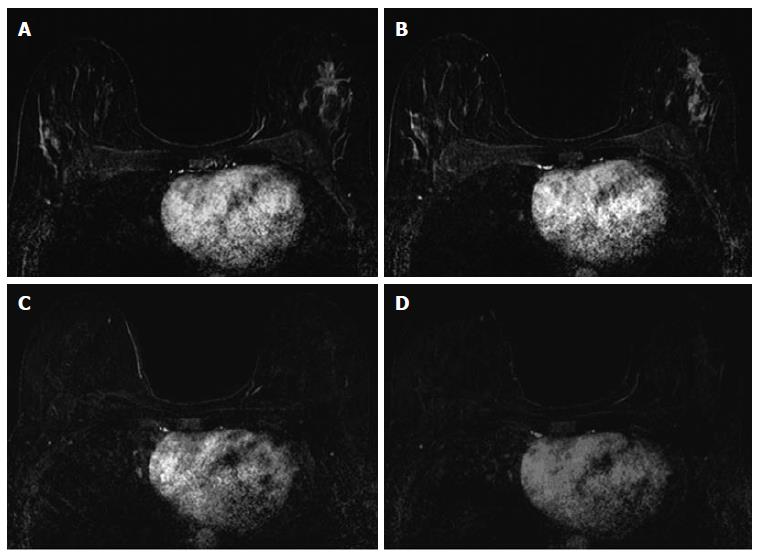
Figure 3 Thirty-seven years old woman with HR+ left breast cancer.
A and B: Baseline axial T1-weighted post-gadolinium fat-saturated magnetic resonance image demonstrates a speculated mass and contiguous non-mass enhancement extending posteriorly for a total of 7 cm of disease in the upper outer breast; C and D: Post-neoadjuvant chemotherapy magnetic resonance imaging demonstrates decrease in size and degree of enhancement of prior findings. Surgical pathology demonstrates 6.9 cm of invasive ductal carcinoma.
- Citation: Price ER, Wong J, Mukhtar R, Hylton N, Esserman LJ. How to use magnetic resonance imaging following neoadjuvant chemotherapy in locally advanced breast cancer. World J Clin Cases 2015; 3(7): 607-613
- URL: https://www.wjgnet.com/2307-8960/full/v3/i7/607.htm
- DOI: https://dx.doi.org/10.12998/wjcc.v3.i7.607


