Copyright
©The Author(s) 2015.
World J Clin Cases. May 16, 2015; 3(5): 474-478
Published online May 16, 2015. doi: 10.12998/wjcc.v3.i5.474
Published online May 16, 2015. doi: 10.12998/wjcc.v3.i5.474
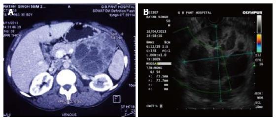
Figure 1 Contrast enhanced computed tomography and endoscopic ultrasound for pancreas.
A: Contrast enhanced computed tomography shows 10.8 cm × 8.1 cm × 9.1 cm solid cystic mass arising from body and tail of the pancreas; B: Endoscopic ultrasound showing a well-encapsulated heteroechoic mass inseparable from tail of pancreas.
- Citation: Gupta G, Saran RK, Godhi S, Srivastava S, Saluja SS, Mishra PK. Composite pheochromocytoma masquerading as solid-pseudopapillary neoplasm of pancreas. World J Clin Cases 2015; 3(5): 474-478
- URL: https://www.wjgnet.com/2307-8960/full/v3/i5/474.htm
- DOI: https://dx.doi.org/10.12998/wjcc.v3.i5.474


