Copyright
©The Author(s) 2015.
World J Clin Cases. Mar 16, 2015; 3(3): 322-326
Published online Mar 16, 2015. doi: 10.12998/wjcc.v3.i3.322
Published online Mar 16, 2015. doi: 10.12998/wjcc.v3.i3.322
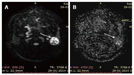
Figure 3 Axial diffusion weighted magnetic resonance imaging.
(A) At b = 1000 s/mm2 hyperintensity is noted throughout the mass (arrow) except the cystic areas. These regions were dark (arrow) on ADC map (B) consistent with restricted diffusion.
- Citation: Jaiswal A, Mridha AR, Nath D, Bhalla AS, Thakkar A. Intraparotid facial nerve schwannoma: A case report. World J Clin Cases 2015; 3(3): 322-326
- URL: https://www.wjgnet.com/2307-8960/full/v3/i3/322.htm
- DOI: https://dx.doi.org/10.12998/wjcc.v3.i3.322


