
















©The Author(s) 2015.
World J Clin Cases. Mar 16, 2015; 3(3): 231-244
Published online Mar 16, 2015. doi: 10.12998/wjcc.v3.i3.231
Published online Mar 16, 2015. doi: 10.12998/wjcc.v3.i3.231
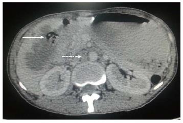
Figure 4 Axial contrast-enhanced computed tomography abdominal section shows ill-defined mass replacing gall bladder fossa with direct infiltration of hepatic segment V.
Cholecystoduodenal fistula (arrow) is noted along with retroperitoneal lymph nodal metastasis (dashed arrow).
- Citation: Dwivedi AND, Jain S, Dixit R. Gall bladder carcinoma: Aggressive malignancy with protean loco-regional and distant spread. World J Clin Cases 2015; 3(3): 231-244
- URL: https://www.wjgnet.com/2307-8960/full/v3/i3/231.htm
- DOI: https://dx.doi.org/10.12998/wjcc.v3.i3.231